2 TRAN et al: DUAL ROLES OF OXOSTEPHANINE AS AURORA KINASE INHIBITOR AND ANGIOGENESIS SUPPRESSOR
during the prophase, prometaphase and metaphase; transfer to the midzone with anaphase onset; and remain in the midbody in telophase and cytokinesis (10). The mislocaliza- tion of any CPC members, including Aurora B, can lead to a defection in mitosis and cytokinesis (10,11). Apart from the pivotal functions in cell division, Aurora A and B kinases are also involved in tumor angiogenesis. These enzymes phosphorylate MYCN, regulate vascular endothelial growth factor (VEGF) production, and inhibit the proliferation and tube formation of human endothelial cells (12-14). Aurora C kinase has been found in cells that undergo meiosis and has a unique physiological role in spermatogenesis (15). The limitation in understanding the role of Aurora C may stem from the high sequence homology between this kinase and Aurora B, leading to the overlapping in the function of these proteins (16). Aurora C can rescue the genetic stability of the cells in case Aurora B is absent (17). Previously, it was demonstrated that the overexpression of Aurora C induces abnormal cell division, resulting in centrosome amplification and multinucleation in cells (17).
The overexpression of Aurora kinases has been observed in a broad range of human solid tumors, such as gliomas, and colorectal, breast, ovarian and pancreatic cancer (18), as well as in liquid tumors such as diffuse large B-cell lymphoma (19). Moreover, Aurora kinases have been found to be associated with genetic instability and aneuploidy in tumors (20). Hence, it is not surprising that Aurora kinases have become attractive targets in cancer treatment. The development of Aurora inhibi- tors has drawn the attention of several scientists from academic institutes and pharmaceutical companies. Over the first two decades of the 21st century, a series of Aurora kinase inhibi- tors were produced, which were Aurora A- or B-selective, or pan inhibitors. Although these compounds exhibit preclinical and clinical efficacy, no Aurora kinase inhibitor has yet been approved for clinical use due to their poor outcomes (18). Thus, there is an urgent need for the identification of novel small molecule inhibitors.
Oxostephanine is a substance belonging to the group of aporphine alkaloids isolated from several plants of the genus Stephania. Previous studies have demonstrated that this substance exerts a potent cytotoxic effect on several cancer cell lines, such as KB (human epithelial carcinoma), HepG2 (human hepatocellular carcinoma), GLC4/Adr (human small cell lung adriamycin-resistant carcinoma), K562 (human chronic myelogenous leukemia) and K562/Adr (human chronic myelogenous leukemia resistant to adriamycin) (21), whereas it has a minimal toxic effect on normal cells (MRC-5; human fetal lung fibroblasts) (22). In addition, oxostephanine has been shown to exhibit potent activity against breast cancer cells and MOLT-3 acute lymphoblastic leukemia cells (21). Moreover, Knockleby et al (23) revealed that oxostephanine inhibited the activity of Aurora kinase A and B by the competition of ATP binding sites in an in vitro kinase assay.
The aim of the present study was to examined the effects of oxostephanine extracted from Vietnamese Stephania dielsiana
Y.C. Wu (S. dielsiana) as a novel Aurora kinase inhibitor on an ovarian cancer cell line (OVCAR-8). As demonstrated herein,
S. dielsiana may prove to be a potent Aurora kinase inhibitor, as well as an anti-angiogenic agent with potential to be devel- oped into an anticancer drug.
Materials and methods
Compound preparation. The stems and leaves of S. dielsiana were collected in Ba Vi District, Hanoi, Vietnam in October, 2019 and identified by the Department of Botany, Hanoi University of Pharmacy, Hanoi, Vietnam. A voucher specimen (no. SD10/2019) has been deposited at the Department of Botany and Pharmacognosy, Vietnam University of Traditional Medicine, Hanoi, Vietnam. The process used for the isolation and characterization of oxostephanine from the leaves of
S. dielsiana in Vietnam has been previously published (22,23). In brief, the leaves of S. dielsiana (7 kg) were extracted with 95% MeOH (3x15 liters, 3 days each) at room temperature. The extracts were concentrated in vacuo to yield a MeOH extract (680 g), which was suspended in H2O (2.5 liters) and adjusted to pH 3 with 10% HCl. The acidic aqueous phase was filtered off. The filtrate was loaded on ion‑exchange resin, eluted with 20% MeOH until the eluate approached colorless to give the nonalkaloid parts, and then eluted with 2% NaOH in 65% MeOH solution (five‑fold of retention volume) to yield the crude total alkaloids. The alkaloid-containing solution was acidified to pH 5 with 10% HCl and partitioned with EtOAc (3x2 liters) to yield the EtOAc extract (65 g).
The EtOAc-soluble portion was subjected to silica gel column chromatography eluted with gradient systems of CH2Cl2-MeOH (100:0, 100:10, 100:30 and 100:50, v/v). The
eluted fractions were evaluated and pooled according to thin
layer chromatography (TLC) analysis, resulting in six major fractions (SDE.1-SDE.6). The purification of SDE.6 over Sephadex LH-20 (100% MeOH) was performed using the same methodology, and subsequent preparative TLC, eluted with CH2Cl2-MeOH (20:1) yielded oxostephanine (8.6 mg). The purification of oxostephanine by repeating recrystallization in a mixture of methanol and ethanol yielded pure oxostephanine compound as an amorphous yellow-orange powder (purity 99.0% as a percentage of the peak area using a HPLC-DA system (Agilent 1260 Infinity II; Agilent Technologies, Inc.).
Cell lines and culture. OVCAR-8 (human ovarian carci- noma-8) and HeLa (Aurora B-GFP) cells were grown in Dulbecco's modified Eagle's medium (DMEM; Gibco; Thermo Fisher Scientific, Inc.). Human dermal fibroblasts (hFBs) were cultured in DMEM/F12 medium (Gibco; Thermo Fisher Scientific, Inc.). The media were supplemented with 10% fetal bovine serum (FBS) (Gibco; Thermo Fisher Scientific, Inc.), 100 units/ml penicillin and 100 µg/ml streptomycin (Gibco; Thermo Fisher Scientific, Inc.). Human umbilical vein endothe- lial cells (hUVECs) were cultured in EBM-2 medium (Lonza Group, Ltd.). Umbilical cord-derived mesenchymal stem cells (UC‑MSCs) were grown on the surface of culture flasks coated by CELLstart™ CTS™ (CELLstart) in StemMACS™ MSC Expansion medium (StemMACS) (Miltenyi Biotec). All the cells were cultured in an incubator at 37˚C with 5% CO2. The hUVECs, hFBs and UC-MSCs were provided by Vinmec Research Institute of Stem cell and Gene Technology, and they were not immortalized cell lines. The protocols for cell isolation were approved by the Ethics Committee of Vinmec International Hospital (Document no. 40/2020/QD-Vinmec for hUVECs and UC-MSCs, signed and dated on December 24, 2020; Document no. 311/2018/QD-Vinmec for hFBs, signed
INTERNATIONAL JOURNAL OF MOLECULAR MEDICINE 50: 133, 20223
and dated on September 11, 2018). The HeLa (Aurora B-GFP) cells were kindly provided as a gift from Professor Stefan Dimitrov at Institute Albert Bonniot (present name is Institute for Advanced Biosciences) (11,24).
Cell viability assay. Cell viability was assessed using sulforho- damine B (SRB) assay. The cells were seeded at a density of 3x10³ cells/well in 96-well plates and incubated with oxostepha- nine for 24, 48 and 72 h at six concentrations differed by five from the highest of 25 to 5, 1, 0.2 and 0.04 µM. Subsequently, the medium was removed, and the cells were stained with 4% SRB (Millipore, Sigma) for 10 min at room temperature after fixing with 10% TCA (MilliporeSigma) for 1 h at 4˚C. The absorbance was measured at 540 nm using a microplate reader (BioTech Power Wave XS; BioTek Instruments, Inc.).
Real-time analysis of cell proliferation using the xCELLigence system. The proliferation assay was performed using the xCel- ligence system (ACEA Biosciences; Agilent Technologies, Inc.). Media (100 µl/well) were added to each 96-well of an E-plate (ACEA Biosciences; Agilent Technologies, Inc.) to take the background reading for 15 min. In the meantime, the cells were resuspended in medium, and 80 µl cell suspension were added to yield a cell density of 3x10³ cells/180 µl/well. Following incubation for 30 min at room temperature, the E-plate was placed into the RTCA SP station in an incubator. After 24 h, the cells were treated with oxostephanine (125, 25, 5, 1 and 0.2 µM) and VX-680 (Vertex and Merck; 25, 5, 1,
0.2 and 0.04 µM). Dynamic cell proliferation was monitored in 30-min intervals from the seeding point till the end of the experiment with a total of >200 h. The electrical impedance was measured using RTCA-integrated software of the xCEL- Ligence system as a dimensionless parameter termed cell index (CI). Normalized CI values were used to obtain the IC50 values, doubling times and other evaluations.
Immunofluorescence. The cells were grown on glass coverslips for 24 h before being treated with either oxostephanine (5 µM) or VX-680 (0.2 µM) with or without paclitaxel (0.035 µM; Millipore, Sigma) and incubated for 15 h in an incubator at 37˚C with 5% CO2. Paclitaxel was used to synchronize the cells to the M phase in the cell cycle, in order to obtain dividing cells. The cells were then fixed with 4% paraformal- dehyde and 2% sucrose for 15 min at 37˚C, permeabilized with 0.2% Triton X-100 for 10 min, blocked with 5 mg/ml BSA, and incubated with primary antibodies for 2 h at room temperature. Phosphorylated histone H3 was detected using a polyclonal rabbit antibody (ab183626, Abcam), at a dilution of 1:500. Aurora B was detected using mouse monoclonal antibodies (36-5200, Invitrogen; Thermo Fisher Scientific, Inc.), at a dilution of 1:250. DNA was visualized with 5 µg/ml Hoechst 33342 (Invitrogen; Thermo Fisher Scientific, Inc.) or 2 µg/ml propidium iodide (PI; Thermo Fisher Scientific, Inc.). Images were collected using a ZEISS 510 Laser Scanning Confocal (LSM) microscope with 40X or 63X objectives (Carl Zeiss AG). For the HeLa (Aurora B-GFP), the cells were grown on a Lab-Tek chamber coverglass (Nalge Nunc International). Following 24 h of treatment with the compounds at concentra- tions of oxostephanine (5 µM) or VX-680 (0.2 µM), cells were observed without fixing.
As regards the cell nuclear morphological examination, the cells were incubated with either oxostephanine (5 µM) or VX‑680 (0.2 µM) for 48 h. The cells were then fixed with 4% paraformaldehyde and 2% sucrose for 15 min at 37˚C, permea- bilized with 0.2% Triton X-100 for 10 min and stained with 5 µg/ml Hoechst 33342. Following incubation for 15 min, the cells were collected, washed with phosphate-buffered saline (PBS; Millipore, Sigma), and observed using a LSM micro- scope. Images were analyzed using LSM Image Browser (Carl Zeiss AG).
Apoptosis assay. Apoptosis assay was performed using the Alexa Fluor 488 Annexin V/dead cell apoptosis kit (Invitrogen; Thermo Fisher Scientific, Inc.). As mentioned in the kit, Annexin V is a phospholipid binding protein, and it specifically binds to negatively charged phosphatidyl- serine molecules exposure on the surface of apoptotic cells. Following treatment of the cells with either 0.5 µM oxostepha- nine or 0.2 µM VX-680 for 48 h, the cells were harvested and prepared for apoptosis analysis. Briefly, the cells were washed with PBS, then suspended in Annexin-binding buffer to obtain a density of 106 cells/ml. The cell solution was then incubated with 5 µl Alexa Fluor® 488-Annexin V and 100 µl PI working solution for 15 min at room temperature. Subsequently, 400 µl Annexin-binding buffer were gently mixed into the solution with and the cell solution was analyzed on a FACS Canto II System (BD Biosciences). For the visualization of apoptotic marker expression, following 24 h of treatment with the compounds, the cells were incubated with Alexa Fluor® 488-Annexin V for 30 min and observed under a LSM micro- scope.
Multicellular tumor spheroid assay. OVCAR-8 spheroids were created using the hanging drop method as previously described (25). A total of 15 µl of the medium that contained 5x10³ cells were added to each circle on the inverted cover of a 96-well plate to create one spheroid. The cover was then placed upside down on the plate coated with sterile agarose 1.5% (w/v) containing 200 µl complete medium. Following 48 h of incubation in a humidified chamber with 5% CO2 at 37˚C, spheroids were transferred from the cover into each well of the agarose-coated plate and further cultured in DMEM (Gibco; Thermo Fisher Scientific, Inc.) supplemented with 10% FBS (Gibco; Thermo Fisher Scientific, Inc.). Spheroids were treated with oxostephanine under two conditions: i) The compound was added to the cell preparation before making the hanging drop; and ii) the compound was added after transferring the formed spheroids into the culture wells. Two concentrations at 5 and 1 µM of Oxostephanine were used in both conditions. Images were obtained using an Axiovert 40CFL microscope (Carl Zeiss AG) with Powershot G9 camera. These images were analyzed using Axio version 4.5 software (Carl Zeiss AG) to determine the spheroid diameter. The approximated volume (V) of each spheroid was calculated as follows: V= (4/3) x π x (D1/2) x (D2/2)2, where D1 and D2 were the longest and shortest diameters, respectively (26).
RNA extraction and reverse transcription-quantitative PCR (RT-qPCR). Total RNA was extracted from the five cell lines using the RNeasy Mini kit (Qiagen GmbH) according to the
4 TRAN et al: DUAL ROLES OF OXOSTEPHANINE AS AURORA KINASE INHIBITOR AND ANGIOGENESIS SUPPRESSOR
Table I. Sequences of specific primers used for RT‑qPCR.
Accession no. | Primer sequence | Amplicon size (bp) | (Refs.) | |
Aurora A | NM_198433.3 | Fw 5'-TTCCAGGAGGACCACTCTCTGT-3' | 69 | (27) |
Rv 5'-TGCATCCGACCTTCAA TCATT-3' | ||||
Aurora B | NM_001313950.2 | Fw 5'-CGCAGAGAGATCGAAATCCAG-3' | 85 | (28) |
Rv 5'-AGATCCTCCTCCGGTCATAAAA-3' | ||||
VEGF | NM_001025366.3 | Fw 5'-AGGAGGAGGGCAGAATCATCAC-3'; | 90 | (29) |
Rv 5'-ATGTCCACCAGGGTCTCGATTG-3' | ||||
β-actin | NM_001101.5 | Fw 5'-ACAGAGCCTCGCCTTTG-3' Rv 5'-CCTTGCACATGCCGGAG-3' | 110 | (30) |
Có thể bạn quan tâm!
-

 Hình Thái Các Dòng Tế Bào Dưới Tác Dụng Của Sd1, Sd2, Sd3, Sd4, Sd5 Tại Thời Điểm 48H (Vk 10X, Zoom 5,6)
 -
 Nghiên cứu thành phần hóa học và đánh giá tác dụng kháng ung thư của thân lá cây củ dòm Stephania dielsiana Y.C. Wu - 40
 -
 Nghiên cứu thành phần hóa học và đánh giá tác dụng kháng ung thư của thân lá cây củ dòm Stephania dielsiana Y.C. Wu - 41
 -
 Nghiên cứu thành phần hóa học và đánh giá tác dụng kháng ung thư của thân lá cây củ dòm Stephania dielsiana Y.C. Wu - 43
 -
 Nghiên cứu thành phần hóa học và đánh giá tác dụng kháng ung thư của thân lá cây củ dòm Stephania dielsiana Y.C. Wu - 44
 -
 Nghiên cứu thành phần hóa học và đánh giá tác dụng kháng ung thư của thân lá cây củ dòm Stephania dielsiana Y.C. Wu - 45
Xem toàn bộ 368 trang tài liệu này.
Fw, forward; Rv, reverse.
manufacturer's instructions. A total of 1 µg total RNA from each sample was converted into cDNA using the M-MLV cDNA Synthesis kit (Enzynomics, Inc.). The reaction was performed at 25˚C for 10 min, 42˚C for 60 min, 95˚C for 5 min, and held at 4˚C on a SimpliAmp™ Thermal Cycler (Applied Biosystems; Thermo Fisher Scientific, Inc.). The cDNA products from each sample were used to perform qPCR. A total of 1 µl five‑time diluted cDNA was used for qPCR, and reagents were mixed followed by PCR using the SensiFAST SYBR® Lo-ROX kit (Bioline Pty Ltd, Meridian Bioscience, Inc.). The primers used are listed in Table I. β-actin mRNA was used as an internal control gene to normalize the data. RT‑qPCR was performed for the initial activation at 95˚C for 20 sec, followed by 40 cycles at 95˚C for 10 sec, 63˚C for 30 sec, and 70˚C for 1 sec. The melting curve was analyzed using the instrument default setting. The assays were performed in trip- licate on a Light Cycle® 96 system (Roche Diagnostics). The DDCq method (31) was used for the quantification of mRNA expression.
Wound healing assay. The hUVECs and hFBs were cultured in EGM-2 endothelial cell growth medium-2 Bulletkit (Lonza Bioscience) and DMEM/F12 (Gibco; Thermo Fisher Scientific, Inc.) supplemented with 10% FBS, respectively, to reach the completed confluence in 24‑well plates. The cells were then supplemented with mitomycin C (5 µg/ml) to inhibit cell proliferation. Thereafter, the cells were cultured in serum-free medium for 24 h (hUVECs) and 48 h (hFBs). Scratches were created using cell scrapers SPLScar (SPL Life Sciences Co., Ltd.), and floating cells were removed by washing the wells twice with PBS. Oxostephanine was incubated with the cells at three concentrations of 25, 5 and 1 µM for 24 h (hUVECs) and 48 h (hFBs). Images were captured every 6 h (Olympus IX73 Inverted Microscope, Olympus Corporation) from the scars created. The cell migration ability was analyzed using ImageJ software (version 1.53e, National Institutes of Health).
Colony formation assay. The hUVECs and hFBs were seeded in a six-well plate at a density of 1x10³ cells/well and treated with oxostephanine at four different concentrations (25, 5, 1 and 0.2 µM) for 24 h. The medium was refreshed, and the cells were then incubated in a humidified incubator with 5% CO2 at 37˚C for a further 10 days. The cells were then stained with
Giemsa (Millipore, Sigma) for 5 min at room temperature after fixing with 70% methanol for 10 min at room temperature. The formation of colony units of endothelial cells (CFU-ECs) and fibroblasts (CFU‑Fs) was observed, photographed and counted using an Axiovert 40 Inverted Microscope (Carl Zeiss AG) (magnification, x4). The number of colonies was determined per 1,000 cells at seeding.
Growth factor analysis using luminex assay. Growth factors, including VEGF‑A, fibroblast growth factor‑2 (FGF‑2) and hepatocyte growth factor (HGF), were analyzed using Luminex assay with ProcartaPlexTM Multiplex Immunoassays (Human Custom ProcartaPlex 4‑Plex kit; Thermo Fisher Scientific, Inc.). The conditioned media was prepared by culturing cells to 90% confluency in an appropriate medium without supple- ment or FBS for 48 h. The conditioned medium was then collected and kept on ice prior to use. Reagents and procedures were processed following the manufacturer's instructions. The luminescent signals of the growth factors were detected using a LuminexTM 100/200TM system equipped with the xPONENT 3.1 software (Luminex Co., Ltd).
Tube formation assay. The tube formation assay was performed using Angiogenesis Assay kit (ab204726, Abcam). Briefly, extracellular matrix solution (Matrigel, supplied with the kit, Abcam) was added to a 96-well plate and incubated for 1 h at 37˚C to allow the solution to form a gel. hVUECs were seeded at 1.5x10⁴ cells/well (three replicates per group) on the gel and incubated with oxostephanine at two concentrations of 5 and 1 µM. For the background control wells, no Matrigel was added. Suramin (supplied with the kit, Abcam) was used as an angiogenesis suppressor control. Following 8 h of incubation at 37̊C in the incubator, the tube formation was examined using an inverted microscope. The total tube length, total branching points and mean tube length were analyzed using Wimasis software (Web-based version, wimasis.com).
Statistical analysis. All statistical analyses were performed using R software version 3.4.4. The differences between groups were assessed using an unpaired t-test, two-way ANOVA and Tukey's HSD tests. A P-value <0.05 was considered to indicate a statistically significant difference. All data are presented as the mean ± SD.
INTERNATIONAL JOURNAL OF MOLECULAR MEDICINE 50: 133, 20225
Results
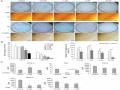
Real-time analysis of the effects of oxostephanine on OVCAR-8 cancer cells. The present study performed a cyto- toxicity analysis of oxostephanine using the OVCAR-8 cell line with the xCELLigence RTCA system. During >200 h of incubation, the viability, number, morphology and adherent ability of the cells were recorded and visualized as a graph (Fig. 1A). The utilities in the RTCA Control Unit software allowed for the creation of a dose-response curve and the calculation of the IC50 value of the drug at different time points. The results revealed that oxostephanine and VX-680 exerted a similar effect on cell proliferation; the higher concentrations of the compounds the greater the inhibitory effects on cell growth. In the control wells, the cell index values gradually increased and peaked at the time point of 140 h, with a CI of 32 (Fig. 1A). In the wells treated with the two highest concentrations of 125 and 25 µM oxostephanine, cell proliferation was entirely inhibited compared to the control with the CI values decreasing after 3 h of incubation, indicating that the cells could not grow, but were killed. At the oxostephanine concentration of 5 µM, the cell proliferation rate was approximately half that of the control, with the time to get the peak of CI values was 165 h. At the oxostephanine concentration of 1 µM, the peak was reached at the same time but with a smaller value equivalent to 78% of the control. At the smallest concentration of 0.2 µM oxostephanine, the cell proliferation was lower than that of the control. For the wells treated with VX-680, while all cells were killed at the two highest concentrations, the CI values at the peaks associated with the other concentrations were smaller and were observed at later time points than those of the control (Fig. 1A). Using RTCA software, the IC50 values at different time points of incubation from 24 to 120 h were calculated. The IC50 values were from 3.8-7.3 µM for oxostephanine and 0.2-0.6 µM for VX-680 (Table II).
The doubling time of the OVCAR-8 cells was also affected by these two compounds. Following treatment with VX-680, the cells did not grow and died rapidly following the addi- tion of the substance expressed by the minus values of the doubling time at the three highest concentrations. In terms of oxostephanine, the majority of the doubling time was higher compared to the controls, indicating that the proliferation of cells was inhibited (Fig. 1B). Of note, a change in the size of the cells treated with oxostephanine and VX-680 at low concentrations was observed. The cells increased their size following the incubation time. Not only the cell size, but the immunostaining of these cells also indicated that there was a significant increase in the nuclei area (Fig. 1C). Additionally, the morphology of the cell nuclei was changed, with the nuclei becoming heterogeneous, multi-lobed and enlarged, that were not homogeneous or oval-shaped as in the controls (Fig. 1C). Using the LSM image browser software, the nuclear area was measured. The data indicated that the nucleic size of the cells treated with oxostephanine or VX-680 was three-fold larger than that in the control group (Fig. 1D). Taken together, these results demonstrated that oxostephanine inhibited the proliferation of OVCAR-8 cells in the micromolar range. The real-time effects of oxostephanine were comparable to those of VX-680, an Aurora kinase inhibitor.
Apoptosis induction is a characteristic of Aurora kinase inhibitors (18,23). Hence, the present study examined whether oxostephanine could induce the apoptosis of OVCAR-8 cancer cells. At the oxostephanine concentration of 5 µM, we observed the expression of phosphatidylserine molecule, an apoptosis marker, that binding to Annexin-V on the cell surface after 24 h of incubation (Fig. 1E). The rate of cells positive with Annexin-V was calculated from the sum of Q1-1 (early apoptosis) and Q2-1 (late apoptosis) quadrants in the flow cytometry plots (Fig. 1F). Accordingly, the percentage of oxostephanine (5 µM)-treated cells positive for Annexin-V was 30.4±6.8%, which was 7.4-fold higher than that of the control (4.1±0.8%). Moreover, a 33.7±5.1% cell population was positive for Annexin-V when treated with 0.2 µM VX-680 (Fig. 1F).
Oxostephanine inhibits the growth of OVCAR-8 spheroids. The effects of oxostephanine on the growth of OVCAR-8 cells in 3D culture were investigated. When adding the substance at the time of spheroid preparation, this compound prevented 70% spheroid formation at 5 µM and 58% spheroid forma- tion at 1 µM. A similar result was obtained with VX-680; only 22.5% of spheroids could be formed at the concentration of 0.2 µM (Fig. 2A). Moreover, the volume of the formed spheroids was smaller than that of the control (Fig. 2B). After transferring the spheroids into agar plates, the growth was unaltered at the concentration of 1 µM, whereas this decreased at the concentration of 5 µM following the time of culture even with the absence of the compound (Fig. 2C). For the other treatments, oxostephanine was added and maintained in the medium after the spheroids were transferred into the agar plate. Under this condition, after 7 days, the substance inhib- ited the growth of spheroids, with the size decreasing 4.3-fold at 5 µM and 2.7-fold at 1 µM. The effect of oxostephanine on spheroid growth was even more prominent than that of VX-680 at 0.2 µM, with a decrease of 2.1-fold in the volume on day 7 of treatment. Moreover, the control increased the spheroid volume 3-fold on day 7 of culture on agar (Fig. 2C). Furthermore, the morphology of the treated spheroids was also changed into loose cell clusters with numerous cells separately surrounded, in contrast to the tight and impact control spheroids (Fig. 2D).
Oxostephanine inhibits Aurora kinase expression and activity. To characterize oxostephanine as a novel Aurora kinase inhibitor, the effect of this compound on the phos- phorylation of H3S10ph was evaluated in OVCAR-8 cancer cells. To collect cells at the mitotic phase, the cell population was synchronized by the addition of paclitaxel followed by incubation with oxostephanine and VX-680 at concentrations of 5 and 0.2 µM, respectively. The images revealed that the fluorescence signal of H3S10ph was markedly decreased in mitotic cells incubated with oxostephanine and VX-680, even with or without paclitaxel (Fig. 3A).
In addition, the distribution of Aurora B was affected by these compounds. In mitotic OVCAR-8 cells, this protein was not expressed at the centromere, but was diffused on the whole chromosomes at the metaphase. Moreover, Aurora B presented as bright dots in the centromere in the control cell group (Fig. 3B). Additionally, RT-qPCR revealed that the mRNA
6 TRAN et al: DUAL ROLES OF OXOSTEPHANINE AS AURORA KINASE INHIBITOR AND ANGIOGENESIS SUPPRESSOR
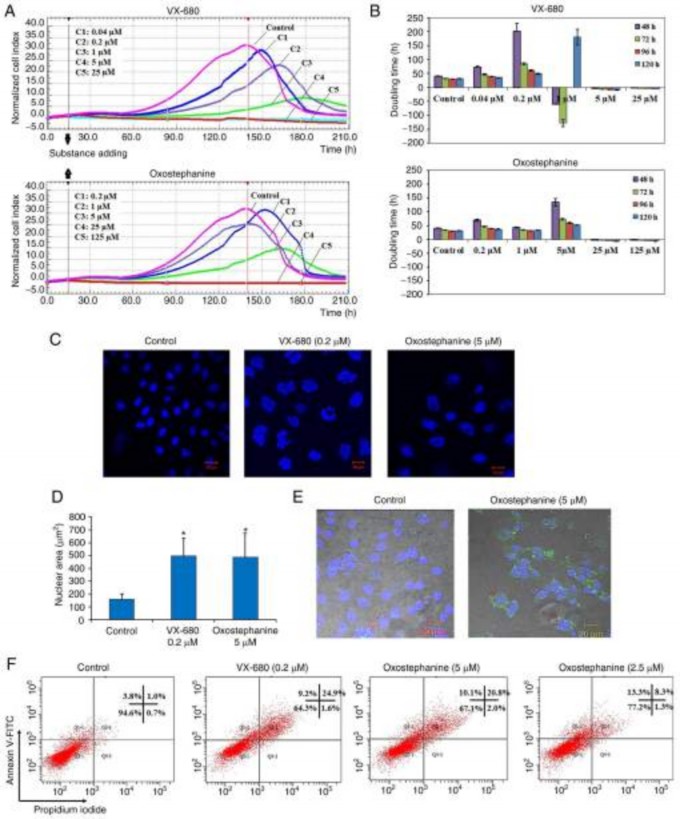
Figure 1. (A) Real-time analysis of OVCAR-8 cell proliferation following treatment with oxostephanine and VX-680. On the plot, the normalized cell index (CI) is shown at 15 h, which is the adding point of the substance. The horizontal axis of the graph was the time of the experiment. (B) Population doubling times of OVCAR‑8 cells were calculated on RTCA system after 48, 72, 96 and 120 h of incubation with the two compounds at five concentrations, as indi- cated in the figures. Of note, the minus values of PDT indicated that the cells died when exposed to the compound at an early stage and no cell growth was counted. (C) Image of cell nuclei following incubation with oxostephanine and VX-680 for 48 h. (D) The average sizes of cell nuclear area were calculated and presented as the mean ± SD. Data were collected from three repeated experiments. (E) Oxostephanine induced the apoptosis of OVCAR-8 cancer cells. Immunofluorescence images of control and oxostephanine‑treated cells stained with Annexin V‑FITC indicated the higher expression of phosphatidylserine molecules on the cell surface in treated cells (green color). (F) Quantitative analysis of the percentage apoptosis in the oxostephanine- and VX-680-treated cells. *P<0.05, vs. control.
INTERNATIONAL JOURNAL OF MOLECULAR MEDICINE 50: 133, 20227
Table II. IC50 values of oxostephanine and VX-680 in OVCAR-8 cancer cells with different incubation times.
Incubation time (h)
------------------------------------------------------------------------------------------------------------------------------------------------------------------------------------
24 | 48 | 72 | 96 | 120 | |
VX-680 (µM) | 0.3±0.02 | 0.6±0.06 | 0.2±0.04 | 0.2±0.07 | 0.2±0.05 |
Oxostephanine (µM) | 7.3±1.5 | 6.6±0.9 | 5.6±0.7 | 4.6±0.8 | 3.8±0.5 |
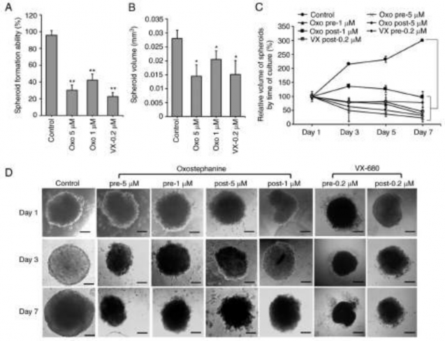
Figure 2. Oxostephanine inhibits the formation and growth of OVCAR-8 spheroids. (A) Spheroid formation in the presence of the compounds. (B) The spheroid volume was reduced following incubation with the compounds. (C) The growth of tumor spheroids was prevented by the two types of treatment: The addition of the compound at the spheroid preparation (pre) and after spheroid formation (post). The days were counted from the time of transferring the spheroid from the hanging drop to the agar plates (day 1, etc.). (D) The morphology of spheroids of cells treated under the two conditions mentioned above. Pre, compounds were added at the time of spheroid preparation; post, compounds were added and maintained in the medium for spheroid growth in the agar plate. Scale bars, 100 µm. *P<0.05 and **P<0.01, vs. control. Oxo, oxostephanine; VX, VX-680.
expression of Aurora B was decreased following incubation with oxostephanine in OVCAR-8 cells (Fig. 3C).
To determine the effects of oxostephanine on the localiza- tion of Aurora B kinase, HeLa cells stably expressing Aurora kinase B-GFP were used. Notably, the diffusion of Aurora B was observed in both living and fixed HeLa cells (Fig. 4). Furthermore, in mitotic cells treated with oxostephanine and VX-680, Aurora B-GFP was observed on the entire chromo- somes when the cells were at metaphase.
In summary, these data illustrated that the treatment of the cells with oxostephanine affected the behavior of Aurora B
during the cell cycle in a similar manner to VX-680, but with
a lower efficiency.
Oxostephanine is selectively cytotoxic on different cell types. The present study also selected three cell lines, including human UC-MSCs, hUVECs and hFBs for the examination of oxostephanine cytotoxicity. Firstly, the expression of Aurora A and Aurora B kinase genes relative to the actin gene control was examined in normal and cancer cells. The results revealed that these genes were highly expressed at the mRNA level, with the highest levels observed in
8 TRAN et al: DUAL ROLES OF OXOSTEPHANINE AS AURORA KINASE INHIBITOR AND ANGIOGENESIS SUPPRESSOR
Figure 3. The phosphorylation of histone H3 at serine 10 and the localization of Aurora B kinase were disrupted in OVCAR-8 cancer cells treated with oxostephanine and VX-680. (A) H3S10ph (green) was suppressed in the presence of oxostephanine (5 µM) and VX-680 (0.2 µM) for 15 h. In the case of synchronization to the pro-metaphase, cells were pre-treated with paclitaxel (0.3 µg/ml) for 8 h, then incubated with the two substances as mentioned above.
(B) Aurora B was deconcentrated on the chromosomal centromeres following treatment with the substances. (C) The expression of Aurora A and Aurora B was decreased at the mRNA level following treatment with oxostephanine and VX-680. *P<0.05, vs. control.
the hUVECs and OVCAR-8 cells, and the lowest in hFBs (Fig. 5A).
Secondly, the cells were incubated with oxostephanine for the analysis of cell death. Following 24 h of incubation with oxostephanine, the death of hUVECs was observed at the two highest concentrations. After 48 and 72 h, the cell death number increased continuously in these wells containing hUVECs. Similar results were detected in UC-MSCs. On the other hand, in the wells of hFBs, no cell death was observed (Fig. 5B). Additionally, the IC50 values were consistent with these observations. The IC50 values from
the hUVECs were 7.9±0.6, 3.1±0.5 and 1.9±0.5 µM after 24, 48 and 72 h of incubation, respectively. However, the IC50 values from the hFBs could not be determined after 24 and 48 h, but were 17.1±0.8 µM after 72 h of incubation. Notably, the cytotoxicity effect of oxostephanine on UC-MSCs was lower than that on hUVECs, but higher than that on hFBs, with IC50 values at 48 and 72 h were 4.7±0.8 and 5.1±0.7 µM, respectively (Fig. 5C). These data, as well as the results of the mRNA levels indicated that the oxostephanine may be more toxic to OVCAR-8 cancer cells and hUVECs, but less on hFBs and UC-MSCs.
INTERNATIONAL JOURNAL OF MOLECULAR MEDICINE 50: 133, 20229
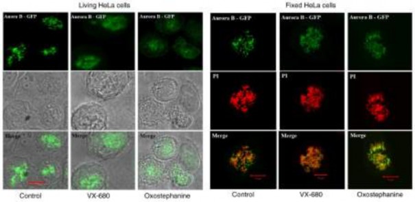
Figure 4. Effects of oxostephanine and VX-680 on the expression of Aurora kinase B in mitotic cells. The Aurora B distribution was determined on living HeLa cells stably expressing Aurora B‑GFP and on fixed HeLa cells. Of note, in the control cells, this protein was located as bright dots on chromosomes at the metaphase; in treated cells, the protein was diffused in the whole chromosomes, particularly in VX-680-treated cells.
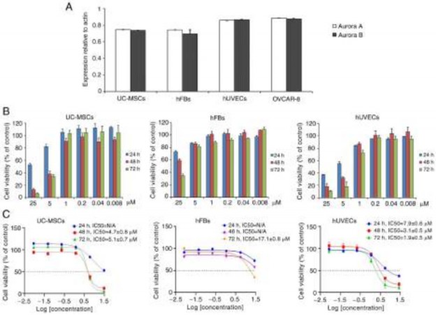
Figure 5. Oxostephanine is selectively cytotoxic to different cell types. (A) mRNA expression of Aurora A and Aurora B kinase in normal and cancer cell lines.
(B) Proliferation of hUVECs and hFBs treated with various concentrations of oxostephanine after 24, 48 and 72 h of incubation. (C) Dose-response growth
inhibition curve for oxostephanine in the three cell types. hUVECs, human umbilical vein endothelial cells; hFB, human dermal fibroblasts.
Oxostephanine reduces colony formation and growth factor secretion by hUVECs and hFBs. The effects of Oxostephanine on the capacity of endothelial progenitor
cells and fibroblast precursor cells to form colonies were then examined. As shown in Fig. 6A, both the number of colonies and the density of cells/colonies were reduced in