1. Introduction
According to the World Health Organization, cancer and cardiovascular disease are the two major causes of mortality worldwide. An overview summarised cancer incidence and mortality rates by sex and age in 2020, for 38 cancer sites in 185 nations and territories worldwide (Ferlay et al. 2021). Numerous disorders, including allergies, oxidative stress, chronic inflammation, cardiovascular diseases and disorders that promote aberrant cell growth, are linked to cancer (Khansari et al. 2009). Although there are a variety of treatment options for cancer, including surgery, chemotherapy and radiotherapy, there is a great deal of research interest in more affordable options using natural ingredients to both prevent and treat cancer (Li et al. 2022, Vinh et al. 2020). Therefore, identification of novel therapeutic components in folk medicines is crucial in the battle against cancer (Duyen et al. 2022, Thang Hoang et al. 2021).
Alkaloids are a large class of organic molecules containing at least one nitrogen atom that exist naturally in both plants and marine organisms. Alkaloids have diverse pharmacological properties, including anti-inflammatory, anticancer, antibacterial and antioxidant properties (Li et al. 2022). Numerous alkaloids have been identified and used in traditional and modern medicine, or have served as the basis for new drug development (e.g. morphine and other opium alkaloids found in opium poppies) (Matos et al. 2022). In addition, berberine, an alkaloid derived from the Berberis genus, has historically been employed in Ayurvedic, Chinese and Middle Eastern folk medicines for its effects against a range of pathogens, including bacteria, viruses, fungi, protozoa and helminths (Kong et al. 2022).
The Menispermaceae plant family, which contains the genus Stephania Lour., is a significant source of medicinal plants (Deng et al. 2011). The alkaloids from this genus are divided into six main groups: hasubanan, aporphine, proaporphine, protoberberine, bisbenzylisoquinoline and morphinandienone (Zhou et al. 2018).
From Stephania dielsiana Y.C.Wu, which is one species of the genus, twenty seven alkaloids were isolated and annouced, such as: sinoacutin, stephanin, ayuthianin, dehydrostephanin, cephamorphinanin, aknadinin, liriodenin, sinomenin, L- tetrahydropalmatin, (-) corydalmin, oxocrebanin, nor-canelillin, crebanin, dehydrocrebanin, stesakin, isolaurelin, oxoputerin, (+)-O-methylbulbocapnin, 8- demethyldehydrocrebanin, vireakin, dehydroisolaurelin, sukhodianin, crebanin N-oxid, và dehydroroemerin, oxostephanin, palmatin, thailandin.
Especially, alkaloids from Stephania dielsiana Y.C.Wu exhibited diverse pharmacological effects, such as cytotoxic, anticancer, anthelmintic and antimicrobial activities (Knockleby et al. 2020, Zhou et al. 2018). As part of an ongoing effort to discover bioactive components from herbal medicine as possible anticancer treatments (Tuan Anh et al. 2021,
Vinh et al. 2019a, Vinh et al. 2019b, Vinh et al. 2020), we describe the structure, extraction, and isolation of two new aporphine alkaloids (1 and 5), along with six known alkaloids (2–4 and 6–8), from the leaves of S. dielsiana. The anticancer properties of isolated compounds were also evaluated by the 3-(4,5-dimethylthiazol-2-yl)-2,5-diphenyl tetrazolium bromide (MTT) protocol using the HepG2, MCF7 and OVCAR8 human cancer cell lines. The results showed that compound 2 exhibited particularly strong cytotoxic activities against HepG2, MCF7 and OVCAR8 cancer cell lines, with IC50 values of 3.20 ± 0.18, 3.10 ± 0.06 and 3.40 ± 0.007 µM, respectively. Furthermore, molecular docking simulations of active compounds were performed to further support our in vitro findings.
2. Results and discussion
2.1. Structure and identification of new compounds
Có thể bạn quan tâm!
-

 Nghiên cứu thành phần hóa học và đánh giá tác dụng kháng ung thư của thân lá cây củ dòm Stephania dielsiana Y.C. Wu - 41
 -
 Nghiên cứu thành phần hóa học và đánh giá tác dụng kháng ung thư của thân lá cây củ dòm Stephania dielsiana Y.C. Wu - 42
 -
 Nghiên cứu thành phần hóa học và đánh giá tác dụng kháng ung thư của thân lá cây củ dòm Stephania dielsiana Y.C. Wu - 43
 -
 Nghiên cứu thành phần hóa học và đánh giá tác dụng kháng ung thư của thân lá cây củ dòm Stephania dielsiana Y.C. Wu - 45
Xem toàn bộ 368 trang tài liệu này.
Dried stems and leaves of S. dielsiana (7.0 kg) were extracted with 95% MeOH (15 L ×3 times) at ambient temperature. MeOH residue was produced after the solvent evaporated under reduced pressure (680 g).
Utilising several chromatographic separation methods, two new alkaloids, stedieltines A–B (1 and 5), and six known alkaloids (oxostephanine (2), oxocrebanine (3), oxostephanosine (4), aristolactam (6) (Achari et al. 1984), crebanine (7) and dehydrocrebanine (8) (Thien et al. 2018)) were purified from the ethylacetate (EtOAc) fraction. Spectroscopic techniques clearly revealed their structures by comparison with previously published data.

Stedieltine A (1) was obtained as a yellowish amorphous powder. It showed a positive reaction to Dragendorff’s reagent, indicating that it was an alkaloid. Its molecular formula, C19H15NO6, with an index of hydrogen deficiency of 13, was defined based on a protonated molecular ion at m/z 354.0979 [M + H]+ (calcd C19H16NO6+, 354.0977) using high-resolution electrospray ionisation mass spectrometry (HRESIMS) (Fig. S9), and from the 13C nuclear magnetic resonance (NMR) data. Analysis of the 1H NMR and 1H-1H COSY spectra (Table S1, Fig S3) revealed the presence of deshielded aromatic proton signals [δH 7.00 (1H, dd, J = 8.0, 1.0 Hz, H-9), 6.81 (1H, t, J = 8.0 Hz, H-10) and 6.67 (1H, dd, J = 8.0, 1.0 Hz, H-11)] characteristizing of an aporphine moiety. A pair of AB doublets [δH 7.81 (1H, d, J = 5.5 Hz, H-4) and 8.28 (1H, d, J = 5.5 Hz, H-5)] were characteristic signals of H-4 and H-5 oxoaporphine derivatives. In addition, two methoxy groups [δH 3.14 (3H, s, 7-OCH3) and 3.83 (3H, s, 8-OCH3)], and one isolated aromatic singlet at δH 7.44 (1H, s), were attributed to H-3, while the singlet at δH 6.14 (2H, s, H-12) was consistent with two hydrogens of a methylenedioxy group in the 1H NMR spectrum. The 13C NMR spectra revealed 19 carbon signals, including 15 aromatic carbons, 1 aromatic O-methyl group [δC 55.9 (8-OMe)], and 1 methylenedioxy [δC 101.9 (C-12)], and a methoxy group [δC 51.2 (7-OCH3)] from one ester [δC 167.0 (C = O)] were also observed.
4
The above data suggested that 1 has an aporphine skeleton and is structurally similar to oxostephanine (2) (Thien et al. 2018), differing only in terms of the replacement of the ketone at C-7 in 2 by a methoxycarbonyl [(δH 3.14/δC 51.2, (OCH3) and δC 167.0 (-COO-)]. This was supported by the present of a methoxy signal at δH 3.14 (3H, s,7- OCH3), correlation from δH
3.14 to δC 167.0 (C-7). An hydroxyl group has been revealed by the chemical signal at δC 145.0 (C-7a), interpreted for an oxygenated aromatic carbon. Additonally, an oxygenated hydrogen signal was demonstrated by a boarden signal on 1H-NMR spectrum at 8.60 ppm in 1. The Heteronuclear Multiple Quantum Correlation (HMQC) data revealed the correlations beetween δH 7.44 (s, H-3) and δC 102.1 (C-3); δH 7.81 (d, J=5.5, H-4) and δC 121.9 (C-4); δH 8.28 (d, J= 5.5, H-5) and δC 139.9 (C-5); δH 7.00 (dd, J=8.0, 1.0, H-9) and δC 111.8 (C-9); δH 6.81 (t, J=8.0, H-10) and δC 118.1 (C-10); δH 6.67 (dd, J=8.0, 1.0, H-11) and δC 123.7 (C-11); δH 6.11 (s, H-
12) and δC 101.9 (C-12); δH 3.14 (s, 3H, 7-OCH3) and δC 51.2 (C-OCH3); The Heteronuclear Multiple Bond Correlation (HMBC) data showed correlations: from δH 6.14 (H-12) to δC 150.0 (C-1), 147.5 (C-2); from δH 7.44 (H-3) to δC 150.0 (C-1), 147.5 (C-2), 135.5 (C-3a), 121.9 (C- 4), and 121.1 (C-6b); δH 7.81 (H-4) to δC 102.1 (C-3), 135.5 (C-3a), 139.9 (C-5), and 121.1 (C- 6b); δH 8.28 (H-5) to δC 135.5 (C-3a), 121.9 (C-4), and 150.2 (C-6a); δH 6.67 (H-11) to δC 112.7 (C-11b), 145.0 (C-7a), and 111.8 (C-9); δH 3.83 (8-OCH3) to δC 147.5 (C-8); δH 7.00 (H-9) to δC145.0 (C-7a), 147.5 (C-8) and 123.7 (C-11) (Figure 2 and S5, S7) further suggested the existence of an aporphine skeleton. The planar structure of 1 was fully assigned and further confirmed by detailed analysis of its two-dimensional NMR spectrums including HMQC, HMBC, 1H-1H COSY (COrrelated SpectroscopY) (Fig. S6) and NOESY (Nuclear Overhauser Effect Spectroscopy) (Fig. S8). Based on its structure, 1 was characterised as a new aporphine alkaloid, named Stedieltine A.
Stedieltine B (5) was obtained as a brown solid, and the molecular formula C17H13NO5 was determined by HRESIMS from the protonated molecular ion observed at m/z 294.0766 [M + H]+ (calcd C17H14NO5+, 294.0766). In the 1H NMR spectrum of 5, the aromatic hydrogen signals at δH 6.95 (1H, dd, J = 8.0, 1.0 Hz, H-9), 7.10 (1H, t, J = 8.0 Hz, H-10) and 7.74 (1H,
dd, J = 8.0, 1.0 Hz, H-11) indicated the existence of a 1,2,3-substituted benzene moiety. Three signals of aromatic singlet hydrogens at δH 6.91 (1H, s, H-3), δH 7.06 (1H, d, J = 5.5 Hz, H-4) and 7.82 (1H, d, J = 5.5 Hz, H-5), as well as the HMBC correlations from H-3 (δH 6.91) to C- 1 (δC 140.8), C-2 (δC 152.7) C-3a (δC 137.3) and C-6b (δC 112.2); H-4 to C-3 (δC 101.2), C-3a
(δC 137.3), C-5 (δC 140.2) and C-6b (δC 112.2); and H-5 to C-3a (δC 137.3), C-4 (δC 115.7) and
C-6a (δC 157.3), revealed the existence of one pentasubstituted and tetrasubstituted benzene moiety. In addition, a singlet downfield hydrogen signal at δH 6.20 (2H, s, 1-OCH2O-2) indicated the presence of a methylenedioxy moiety, and a methoxy group at C-8 by {δH 3.93
5
(s; δC 56.3; -OCH3). The 13C NMR and HSQC spectra of 5 indicated the occurrence of 17 carbons, including 1 sp3 methylene, 15 sp2 carbons and 1 methoxy group. The 15 sp2 carbons could be ascribed to the presence of one double bond and two benzene ring moieties that accounted for 9 of the 12 degrees of unsaturation. The remaining three indices of hydrogen deficiency were assigned to three additional ring systems in 5. The above spectroscopic data analysis revealed that the structure of 5 resembled that of oxostephanine (2) . The difference between 5 and oxostephanine (2) was revealed by comparing the NMR spectral data of 5 (Table
1) with that of oxostephanine, which showed that the carbonyl group at C-7 in oxostephanine was replaced by a bond to an oxygen atom in 5. This assignment was confirmed by the chemical shifts at δC 157.3 (C-6a) and 141.3 (C-7a) as well as by high-resolution electrospray ionisation mass spectrometry (HR-ESI-MS) data. Additionally, HMQC has showed a signal between H-9 (δH 6.95) and C-9 (δC 113.1), that has meant for the lack of one -OCH3 at position 9 in comparing with oxostephanine. Thus, the structure of 5 was determined as shown in Figure 1. The complete and unambiguous NMR assignments of oxostephanine (2), oxocrebanine (3), oxostephanosine (4), aristolactam (6) (Achari et al. 1984), crebanine (7) and dehydrocrebanine
(8) (Thien et al. 2018) were accomplished by comparison of the spectroscopic data (NMR and MS) with literature values.
2.2 Biological evaluation
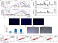
The potential cytotoxicity of compounds 1–8 was examined using three human cancer cell lines: HepG2, MCF7 and OVCAR8 (Tuan Anh et al. 2021). Compound 2 exhibited particularly strong cytotoxic activities against HepG2, MCF7 and OVCAR8 cells, with IC50 values of 3.20 ± 0.18,
3.10 ± 0.06 and 3.40 ± 0.007 µM, respectively (Table 2). Other compounds showed weaker or no significant cytotoxic effects on the human cancer cell lines.
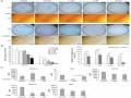
Based on the potential cytotoxic effects of compound 2, a computational study was performed to support the results of the in vitro experiment. The interaction and binding mechanism of active compound 2 with proteins related to cancer were investigated by molecular docking simulations. The results showed that compound 2 had good binding energies of −9.8, −8.0 and
−9.8 kcal/mol for HepG2 (PDB ID: 5EQG), MCF7 (PDB ID: 3ERT) and OVCAR8 (PDB ID:
3OG7), respectively (Figure 3). Furthermore, compound 2 exhibited a hydrogen bond with TRP412 in the active site for HepG2 (PDB ID: 5EQG). Therefore, compound 2 from S. dielsiana is a potential candidate for the development of novel anticancer therapeutic agents. Previously, alkaloids isolated from the genus Stephania were reported to show remarkable anti- inflammatory, antinociceptive and anticancer activities (Deng et al. 2011, Knockleby et al. 2020). In this study, eight compounds (1–8), including two new aporphine alkaloids (1 and 5), were identified in the methanol (MeOH) extract of S. dielsiana by combined column
6
chromatography (CC). Compound 2 may have anticancer effects. Our results suggested that the alkaloids in S. dielsiana might have potential for the treatment of cancer and related diseases.
3. Experimental
3.1. General experimental procedures
The 1H (500 MHz) and 13C NMR (125 MHz) spectra were recorded in deuterated solvents on an AVANCE III HD 500 spectrometer (Bruker, Billerica, MA, USA), operating at 125 MHz for 13C and 400 MHz for 1H. Chemical shifts are reported in ppm (δ) and coupling constants (J) as Hz, relative to those of the solvent signal. Tetramethylsilane (TMS) was used as an internal reference. HRESIMS data were acquired using a 6530 Accurate-Mass Q-TOF LC/MS system (Agilent, Santa Clara, CA, USA). Medium-pressure liquid chromatography (MPLC) was carried using a Biotage-Isolera One system (SE-751 03; Biotage, Uppsala, Sweden). CC was performed using silica gel 65–250 or 230–400 mesh silica gel (Sorbent Technologies, Atlanta, GA, USA), porous polymer gel (Diaion HP-20, 20–60 mesh; Mitsubishi Chemical, Tokyo, Japan), Sephadex LH-20 (Supelco, Bellefonte, PA, USA), octadecyl silica (ODS, 50 μm, COSMOSIL 140 C18-OPN; Nacalai Tesque, Kyoto, Japan) and RP-18 (30–50 μm, YMC*GEL; Fuji Silysia Chemical, Kasugai, Japan). Analytical thin-layer chromatography (TLC) was performed on precoated silica gel 60 F254 (1.05554.0001; Merck, Darmstadt, Germany) and RP-18 F254S plates (1.15685.0001; Merck) and visualised under short wavelength ultraviolet (254 nm) and long wavelength ultraviolet (365 nm). The isolated compounds were visualised by spraying with 10% H2SO4 in water and then heating for 1.5–2 minutes. All procedures were carried out with solvents purchased from commercial sources used without further purification.
Cancer cell lines: HepG2, MCF7 and OVCAR8 cell lines were provided by the American Type Culture Collection (ATCC), then stored in nitrogen liquid in Biology Department, Natural Sciences Univeristy, Hanoi National University. The cells were grown in Dulbecco’s modified Eagle’s medium (DMEM; Gibco; Thermo Fisher Scientific, Inc).
3.2. Identification of plant S. dielsiana
The study materials, which had harvested whole aerial parts from the first branch of trunk of Stephania dielsiana, were collected in Ba Vi district, Hanoi, Vietnam, in October 2019 and a voucher specimen (SD10/2019) was kept at the Department of Botany and Pharmacognosy, Vietnam University of Traditional Medicine, Hanoi, Vietnam.
3.3. Extraction of plant and column chromatography isolation of S. dielsiana
The stems and leaves of S. dielsiana (7 kg) were extracted with 95% MeOH (3 × 15 liters, 3 days each) at room temperature. The extracts were concentrated in vacuo to give a MeOH extract (680 g), which was suspended in H2O (2.5 liters) and adjusted to pH 3 with 10% HCl.
The acidic aqueous phase was filtered off. The filtrate was loaded onto ion-exchange resin, eluted with 20% MeOH until the eluate became colourless to yield the nonalkaloid parts, and then eluted with 2% NaOH in 65% MeOH solution (5× retention volume) to afford the crude total alkaloids. The alkaloid-containing solution was acidified to pH 5 with 10% HCl and partitioned with ethyl acetate (EtOAc) (3 × 2L) to afford the EtOAc extract (65 g).
The EtOAc-soluble portion was subjected to silica gel column chromatography (CC) eluted with gradient systems of CH2Cl2-MeOH (100:0, 100:10, 100:30, 100:50, v/v). The eluted fractions were pooled according to TLC analysis, yielding six major fractions (SDE.1–SDE.6). The SDE.1 fraction was separated by silica gel CC and eluted with n-hexane-acetone (10:1, v/v) to afford 8 (8.8 mg). The SDE.3 fraction was subjected to silica gel CC and eluted with CH2Cl2- acetone (80:1, v/v), using the same methodology as described above for the initial CC of the alkaloid fraction. Subsequent preparative TLC was eluted with n-hexane-acetone (6:1, v/v) to give compound 7 (5 mg). Compounds 1 (5.5 mg) and 4 (3.9 mg) were obtained from fraction SDE.4 by silica gel CC (CH2Cl2-acetone, 18:1) and further separated by RP-18 (MeOH-H2O, 1:2). Purification of SDE.6 over Sephadex LH-20 (100% MeOH), using this same methodology followed by preparative TLC, and eluted with CH2Cl2-MeOH (20:1), yielded compounds 2 (8.6 mg), 3 (2.8 mg) and 5 (3.5 mg). Finally, compound 6 (2.3 mg) was separated from fraction SDE.8 by chromatography on a Sephadex LH-20 column, using MeOH as the eluent, and further isolated and purified by silica gel CC (CH2Cl2-MeOH (10:1).
Stedieltine A (1): yellowish amorphous powder (for 1H and 13C NMR spectroscopic data, see Table S1); HR-ESI-MS m/z 354.0979 [M + H]+ (calcd C19H16NO6+, 354.0977).
Stedieltine B (5): brown solid (for 1H and 13C NMR spectroscopic data, see Table S1); HR-ESI- MS m/z 294.0766 [M + H]+ (calcd C17H14NO5+, 294.0766).
3.4. Cytotoxicity assay
The MTS assay. Cells HepG2, MCF7 and OVCAR8 were seeded at a concentration of 1 x 10⁵ cells/mL, 200 mL/well, into 96-well flat-bottomed tissue culture plates in eight replicates. The MTS assay was carried out using the MTS Cell Proliferation Colorimetric Assay Kit (BioVision, Inc., Milpitas, CA) following the manufacturer’s instruction. In brief, after the cells were cultured with medium for 24 hours, then washed out and replaced with 200 µL fresh warmed the culture medium before adding 20 µL of MTS reagent (3-(4,5-dimethylthiazol-2- yl)-5-(3-carboxymethoxyphenyl)-2-(4-sulfophenyl)-2H-tetrazolium) into each well. The cells were further incubated for 2–4 h at 37oC in standard culture conditions. Then the absorbance was detected at 490 nm with a microplate reader. (Wang Y et al. 2020)
3.5. Molecular docking simulation
The docking study was carried out as described previously (Duyen et al. 2022). Briefly, molecular docking studies were carried out using AutoDock Vina 1.1.2 to determine the binding affinity and interaction of the most active compound (compound 2) with proteins related to cancer, using protocols reported previously (Phong et al. 2022, Phong et al. 2021). The X-ray crystallographic structures of proteins related to cancer, i.e. HepG2 (PDB ID: 5EQG), MCF7 (PDB ID: 3ERT) and OVCAR8 (PDB ID: 3OG7), were obtained from the RCSB Protein Data Bank. Energy minimisation of the active compound was accomplished using Chem 3D Ultra version 20. The molecular docking results were visualised using Discovery Studio 20.1 (Dassault Systemes Biovia, San Diego, CA, USA).
Declaration of Competing Interest
The authors declare that they have no known competing financial interests or personal relationships that could have appeared to influence the work reported in this paper.
Acknowledgments
This research is funded by the Administration of Science Technology and Training – Ministry of Health – Vietnam (according to Decision no. 2721/QD-BYT, dated June 28, 2019, and Contract no. 09/HD-K2DT, dated September 18, 2019).
Appendix A. Supplementary material
Supplementary material related to this article can be found, in the online version, at
References
Achari B, Bandyopadhyay S, Chakravarty AK, Pakrashi SC. 1984. Carbon-13 NMR spectra of some phenanthrene derivatives from Aristolochia indica and their analogues. Org Magn Reson. 22:741-746.
Deng Y, Yu Y, Luo H, Zhang M, Qin X, Li L. 2011. Antimicrobial activity of extract and two alkaloids from traditional Chinese medicinal plant Stephania dielsiana. Food Chem. 124:1556-1560.
Duyen NT, Vinh LB, Phong NV, Khoi NM, Long PQ, Hien TT, Dat NT, Lee KY. 2022. Steroid glycosides isolated from Paris polyphylla var. chinensis aerial parts and paris saponin II induces G1/S-phase MCF-7 cell cycle arrest. Carbohydr Res. 519:108613.
Ferlay J, Colombet M, Soerjomataram I, Parkin DM, Piđeros M, Znaor A, Bray F. 2021. Cancer statistics for the year 2020: An overview. Int J Cancer. 149:778-789.
Khansari N, Shakiba Y, Mahmoudi M. 2009. Chronic inflammation and oxidative stress as a major cause of age-related diseases and cancer. Recent Pat Inflamm Allergy Drug Discov. 3:73-80.
Knockleby J, Pradines B, Gendrot M, Mosnier J, Nguyen TT, Trinh TT, Lee H, Le PM. 2020. Cytotoxic and anti-plasmodial activities of Stephania dielsiana YC Wu extracts and the isolated compounds. Molecules. 25:3755.
Kong Y, Li L, Zhao L-G, Yu P, Li D-D. 2022. A patent review of berberine and its derivatives with various pharmacological activities (2016–2020). Expert Opin Ther Pat. 32:211- 223.
Li C, Wang J, Ma R, Li L, Wu W, Cai D, Lu Q. 2022. Natural-derived alkaloids exhibit great potential in the treatment of ulcerative colitis. Pharmacol Res. 175:105972.
Matos AC, Marques IA, Pires AS, Valentim A, Abrantes AM, Botelho MF. 2022. The potential effect of lidocaine, ropivacaine, levobupivacaine and morphine on breast cancer pre- clinical models: a systematic review. Int J Mol Sci. 23:1894.
Phong NV, Anh DTN, Chae HY, Yang SY, Kwon MJ, Min BS, Kim JA. 2022. Anti- inflammatory activity and cytotoxicity against ovarian cancer cell lines by amide alkaloids and piperic esters isolated from Piper longum fruits: in vitro assessments and molecular docking simulation. Bioorg Chem.106072.
Phong NV, Oanh VT, Yang SY, Choi JS, Min BS, Kim JA. 2021. PTP1B inhibition studies of biological active phloroglucinols from the rhizomes of Dryopteris crassirhizoma: Kinetic properties and molecular docking simulation. Int J Biol Macromol. 188:719- 728.