10 TRAN et al: DUAL ROLES OF OXOSTEPHANINE AS AURORA KINASE INHIBITOR AND ANGIOGENESIS SUPPRESSOR
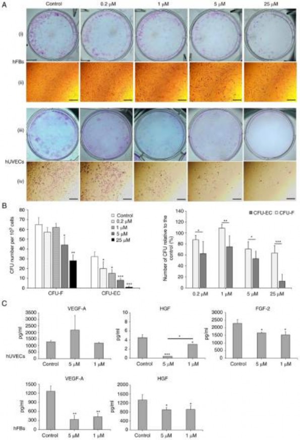
Figure 6. Oxostephanine reduces the colony formation and growth factor secretion by hUVECs and hFBs. (A) Images of CFU-Fs (hFBs) and CFU-ECs (hUVECs) and cell morphology in each type of CFU. CFUs were reduced in both the number of CFU and the number of cells per CFU. (i and iii) Macroscopic images of hFBs and hUVECs culture plates, respectively, following Giemsa staining; (ii and iv) microscopic of a single stained colony in hFBs and hUVECs, respectively. Scale bars, 200 µM. (B) The colony formation ability of hUVECs and hFBs treated with the indicated concentrations of oxostephanine. (C) The secretion of three types of growth factors (VEGF-A, HGF and FGF-2) in the presence of oxostephanine at various concentrations. *P<0.05, **P<0.01 and
***P<0.001, vs. control. hUVECs, human umbilical vein endothelial cells; hFB, human dermal fibroblasts; CFU, colony‑forming units.
INTERNATIONAL JOURNAL OF MOLECULAR MEDICINE 50: 133, 202211
Có thể bạn quan tâm!
-
 Nghiên cứu thành phần hóa học và đánh giá tác dụng kháng ung thư của thân lá cây củ dòm Stephania dielsiana Y.C. Wu - 40
 -
 Nghiên cứu thành phần hóa học và đánh giá tác dụng kháng ung thư của thân lá cây củ dòm Stephania dielsiana Y.C. Wu - 41
 -
 Nghiên cứu thành phần hóa học và đánh giá tác dụng kháng ung thư của thân lá cây củ dòm Stephania dielsiana Y.C. Wu - 42
 -
 Nghiên cứu thành phần hóa học và đánh giá tác dụng kháng ung thư của thân lá cây củ dòm Stephania dielsiana Y.C. Wu - 44
 -
 Nghiên cứu thành phần hóa học và đánh giá tác dụng kháng ung thư của thân lá cây củ dòm Stephania dielsiana Y.C. Wu - 45
Xem toàn bộ 368 trang tài liệu này.
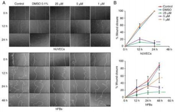
Figure 7. Oxostephanine inhibits the migration of hUVECs and hFBs. (A) Images of cell migration toward the gap in the presence of oxostephanine at the indicated concentrations. (B) Quantitative analysis of gap covering (%) after a time of cell migration. Experiments were repeated in triplicate and data are presented as the mean ± SD. *P<0.05 and ***P<0.001. hUVECs, human umbilical vein endothelial cells; hFB, human dermal fibroblasts.
the treated wells compared to the controls. The numbers of CFU‑ECs and CFU‑Fs were significantly decreased with the two highest concentrations (25 and 5 µM) (P<0.01). Colony formation was also disrupted with the lower concentra- tions of oxostephanine, with a smaller number of colonies compared to the control in hUVECs (P<0.05) (Fig. 6B, left panel. In addition, the inhibitory effects of oxostephanine on colony formation were more prominent in hUVECs than in hFBs, with a smaller number of CFUs relative to the control (%) in the endothelial cells compared to that in the fibroblasts (P<0.05) (Fig. 6B, right panel).
Three types of growth factors, including VEGF-A, FGF-2 and HGF, were measured in the cell culture medium after treating the cells with oxostephanine at 1 and 5 µM. The data indicated that the secretion of these proteins was differed between the cell types. In the controls, both the hUVECs and hFBs secreted HGF with values of 4.5, and 1,333±243.2 µg/ml, respectively (Fig. 6C). Additionally, both the hFbs and hUVECs produced VEGF-A into the medium at a concentration of around ~1,270 pg/ml. The hUVECs secreted a high amount of FGF-2 (2,285.8±240.1 pg/m). Following incubation with oxostephanine, the capacity of growth factor secretion by the cells was consistent with the control regarding the factor component that only hUVECs could secrete all three factors (VEGF-A, HGF and FGF-2) and hFBs secreted only VEGF-A and HGF. However, the amount of all tested growth factors decreased (P<0.05), apart from VEGF-A secreted by hUVECs treated with 5 µM oxostephanine (Fig. 6C). These results demonstrated that oxostephanine affected the secretion of growth factors by cells.
Oxostephanine inhibits the migration of hUVECs and hFBs. Fibroblast and endothelial cell migration is a critical step in the wound healing and angiogenesis processes (32). Thus, in the present study, a wound healing assay was performed to examine the capacity of oxostephanine to regulate the migration of endothelial cells and fibroblasts. In the control group, both hUVECs and hFBs expressed their ability to migrate to close the gap at a more rapid rate; the hUVECs exhibited a greater migratory ability (covering 100% of the wound after 24 h) compared to the hFBs (covering 46.1% of the wound after 24 h) (Fig. 7). When the cells were treated with oxostephanine, a significant decrease in the migration of hUVECs and hFBs were observed (P<0.05; Fig. 7). As regards the hUVECs, the percentage of the wound covered by cells treated with oxostephanine at the concentrations of 25 and 5 µM was ~11% compared to 100% of that in the control group after 24 h, which indicated that the compound inhibited the migration of hUVECs >10-fold (Fig. 7). This inhibitory effect was less prominent in hFBs at the two highest concentrations (5.7-fold decrease at 25 µM and 3.2-fold decrease at 5 µM at 48 h). However, at the concentration of 1 µM, the compound exerted more prominent inhibitory effects on the migration of hFBs than that of hUVECs. These results demonstrated that oxostephanine significantly inhibited the migration of hUVECs and hFBs.
Oxostephanine suppresses angiogenesis in vitro. The effect of oxostephanine on the angiogenesis of hUVECs was examined using tube formation assay. As shown in Fig. 8A, the hUVECs formed a capillary-like network on the Matrigel, with the
12 TRAN et al: DUAL ROLES OF OXOSTEPHANINE AS AURORA KINASE INHIBITOR AND ANGIOGENESIS SUPPRESSOR
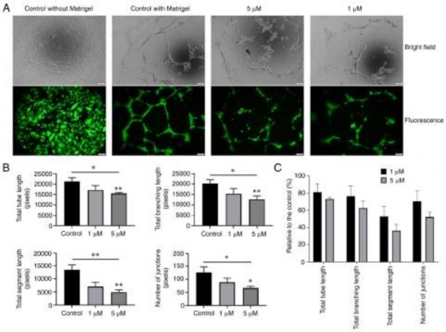
Figure 8. Effect of osxostephanine on the tube formation assay of hUVECs. (A) Representative images and (B) quantification of the tube formation when seeding hUVECs on Matrigel in the presence of the test compounds. (C) Tube formation capacity relative to the control (%) of hUVECs incubated with oxostephanine. Experiments were repeated in triplicate and data are presented as the mean ± SD. *P<0.05 and **P<0.01.
highest number of total tube lengths and tube branching points after 10 h. By contrast, the tube-formation capacity significantly decreased when the cells were treated with 5 µM oxostephanine (P<0.05) (Fig. 8B). The total tube length, tube branching, tube segments and the number of junctions were 72.9±2.1, 62.5±8.4, 36.4±7.2, and 52.1±5.6%, respectively,
compared to the control group. The majority of hUVECs clus- tered, and very few tube-like structures were observed. When the cells were treated with 1 µM oxostephanine, the percentage of total tube length, branching, segments, and number of junc- tions reached 80.8±10, 76.2±12, 52.7±12.2, and 70.3±12.3%,
respectively, compared to the control (Fig. 8C). These findings
suggested that oxostephanine suppressed angiogenesis in vitro.
Discussion
The crucial role of Aurora kinases, particularly Aurora A and B, in cell division, as well as the overexpression of these kinases in a wide range of cancers, renders them a potential target in cancer treatment (18). Oxostephanine extracted from the Stephania plant was first reported by Makarasen et al (21) for its activity in inhibiting the growth of a variety of cancer cell lines. The present study first aimed to characterize oxostephanine, extracted from S. dielsiana leaves in Vietnam, as a novel Aurora inhibitor by comparing the real-time
effects of this substance on cancer cells to those of VX-680, a well-known Aurora kinase inhibitor (33). An ovarian cancer cell line (OVCAR-8), was used to examine the effects of oxostephanine, since Aurora kinase has been reported to be overexpressed in epithelial ovarian cancer, in addition to two recent clinical trials that have used Aurora kinase inhibitors to treat ovarian cancer (34-36). In the present study, the analysis using the xCelligence system revealed similar responses of the OVCAR-8 cells to both compounds (oxostephanine and VX-680) in real-time growth dose-response curves, cell popu- lation doubling time and cellular size change.
Of note, at low tested concentrations of oxostephanine (<5 µM) and VX-680 (1 µM), the cells became aneuploidy with an increase in their size, but not in their number. Previous research has indicated that when Aurora kinase activity is inhibited, the mitotic SAC is activated, which leads to mitotic arrest. However, this SAC could be overridden, which causes the mitotic slippage of cells in the presence of Aurora kinase inhibitors. This phenomenon eventually led to cells becoming aneuploidy or apoptotic (37). In the present study, OVCAR-8 cells treated with oxostephanine and VX-680 at low concentra- tions expressed enlarged and lobed nuclei. Moreover, as shown by immunofluorescence assay, both compounds downregu- lated the phosphorylation of protein histone H3 at serine 10 in cancer cells. These data are consistent with those of the study
INTERNATIONAL JOURNAL OF MOLECULAR MEDICINE 50: 133, 202213
by Knockleby et al (23), demonstrating that oxostephanine inhibited H3S10ph in HeLa cells (23). As the phosphorylation of histone H3 at serine 10 is considered a marker of activated Aurora B kinase (7,8), hence oxostephanine could prevent the function of this kinase.
Previous studies have indicated that the activity of Aurora B is associated with auto-phosphorylation and centro- mere distribution (5,23). Under normal conditions, Aurora B must concentrate at the kinetochore to phosphorylate some proteins in the conserved outer kinetochore KNL1/Mis12 complex/Ndc80 complex (KMN) network, which plays a role in the kinetochore-microtubule attachment (4-6). The present study demonstrated that oxostephanine affected the normal localization of Aurora B kinase; thus, it may inhibit the auto-phosphorylation activity of this enzyme. In the presence of oxostephanine and VX-680, Aurora B diffused to all chro- mosome arms and in the cytoplasm. This phenomenon ocurred in all fixed and living OVCAR‑8 and HeLa cells. By observing living HeLa cells that express Aurora B-GFP, it was noted that the cells that have chromosomes with diffused Aurora B remained longer in metaphase and eventually became aneu- ploidy. This phenomenon of Aurora B has been mentioned with the other inhibitors (24,25). This could be explained by the fact that Aurora B did not concentrate at the kinetochore, leading to an effect on the correct attachment of the chromo- some to the microtubule and subsequently activating the SAC, consequently leading to mitotic slippage, as discussed above. Moreover, oxostephanine decreased the expression of both Aurora A and Aurora B at the mRNA level as did VX-680. The reduction in the levels of these proteins contributed to defects in cell division functions. Taken together, these data demon- strated that oxostephanine was an Aurora kinase inhibitor, and this compound was cytotoxic to OVCAR-8 cells in both monolayer culture and tumor spheroids. It is worth noting that Knockleby et al (23) indicated the effect of Oxostephanine on both Aurora A and Aurora B in the kinase assay. The present study first focused on Aurora B in OVCAR‑8 cells. In future studies, the authors aim to continue to test the effects of oxostephanine on Aurora A kinase in cell culture.
Cancer‑associated mesenchymal stem cells and fibroblasts have been proven to facilitate tumor progression. Recent research has revealed the function of mesenchymal stem cells in glioblastoma resistant to Aurora kinase inhibitor, leading to the recurrence of tumors (38). In acute myeloid leukemia (AML), one mechanism of mesenchymal stem cells used to protect leukemic cells from chemotherapeutic agents is activating Aurora A by increasing IL-6 secretion (39). In a co‑culture system, fibroblasts have been shown to induce the upregulation of Aurora A in non-small cell lung carcinoma to protect the cancer cells from gefitinib treatment (40,41). Fibroblasts can be activated by Aurora B through Wilms tumor 1 signaling, leading to an induction of fibrogenesis (42). Moreover, the downregulation of Aurora B stimulates cellular senescence in hFBs (43). Aurora kinase and stromal cells exert synergistic effects on the development of cancer cells. Moreover, angiogenesis is necessary for the progres- sion of tumors (44). Hence, in the present study examined the effects of oxostephanine on four cell types (UC-MSCs, hFBs, hUVECs and OVCAR-8). Firstly, it was found that all tested cells highly expressed Aurora A and B, with the highest
expression level observed in OVCAR-8 cells and hUVECs, followed by UC‑MSCs, and finally hFBs. Accordingly, the IC50 values of oxostephanine in these cell lines were the lowest in the OVCAR-8 cells and hUVECs, higher in the MSCs, and highest in the hFBs. Moreover, the reduction in the colony-forming units indicated that oxostephanine could inhibit the proliferation of endothelial progenitor cells and fibroblasts. One limitation of the present study was that the presentation of colonies needed improvement as the location of the closely clustered colonies could not be seen. However, the number of colonies could still be counted. At the concen- tration of 5 µM, oxostephanine significantly inhibited the colony formation of hUVECs; however, the colony-forming inhibitory effect was less prominent in hFBs (~30% CFUs). Additionally, in the wound healing assay, oxostephanine also exerted a more prominent inhibitory effect on the migration of hUVECs than hFBs. These results demonstrated the selec- tive activity of oxostephanine toward hUVECs. The targeting of the compound to different cell types may result from the expression of Aurora kinase in these cells. Higher levels of Aurora kinase are associated with a more prominent effect of oxostephanine on the cells. Apart from cell growth, the function of hUVECs in angiogenesis was also disrupted by oxostephanine. These cells could not successfully form tubes in Matrigel in the presence of 5 µM oxostephanine. The anti-angiogenic effect of Aurora kinase inhibitors has been previously reported (13) through their involvement in a signaling pathway that enhances angiogenesis (45) and stabi- lizes N-Myc, which is a well-known oncogene (46,47). These results indicate that oxostephanine functions as a suppressor of angiogenesis.
Furthermore, the data indicated that oxostephanine decreased the production of VEGF-A, HGF and FGF-2, which functions in the proliferation, migration and tube formation processes (48-51), by both hUVECs and hFBs. Notably, in the present study, in hUVECs, the mRNA expression of VEGF-A in cells treated with oxostephanine was not considerably altered; however, the expression of FGF‑2 was significantly decreased compared to the control. This activity of oxostepha- nine differed from VX-680, which has been shown to inhibit VEGF-A expression (13). Nonetheless, the decrease in the levels of FGF‑2 and HFG was sufficient to inhibit the growth and function of hUVECs.
Of note, the effects of oxostephanine one growth factor secretion by cells have not yet been clarified. In addition, the involvement of Aurora kinases in angiogenesis have not yet been elucidated. However, it can be hypothesized that Aurora kinase inhibitors, such as oxostephanine, are cytotoxic toward ovarian cancer cells and endothelial cells, which leads to the inhibition of tumor angiogenesis. Furthermore, even though this compound was less cytotoxic to the other stromal cells such as hFBs and UC-MSCs, it prevented the cell functions that can result in stromal cells being inefficient in supporting tumor growth. This hypothesis was encouraged by a published study on the antitumor activity of the methanol fractional extraction from
S. dielsiana roots on Swiss mice bearing Sarcoma-180 tumors, which reported a 4-fold decrease in tumor volume in the treated mine (52). It is necessary to examine the effects of oxostephanine in vivo using animal models transplanted with human tumor cells. The authors aim to perform such experiments in the future.
14 TRAN et al: DUAL ROLES OF OXOSTEPHANINE AS AURORA KINASE INHIBITOR AND ANGIOGENESIS SUPPRESSOR
In conclusion, the findings of the present study indicate that oxostephanine is a potential Aurora kinase inhibitor. It inhibited the proliferation of ovarian cancer OVCAR-8 cells and multicellular tumor spheroids. Moreover, oxostephanine exhibited selective cytotoxicity to normal cells by inducing a high expression of Aurora kinase A and B. Furthermore, this compound downregulated the expression of growth factors, prevented the migration of hUVECs and hFBs, and reduced tube formation. However, further studies are required for oxostephanine to be developed as an anticancer drug. This compound needs to be tested on other ovarian cancer cell lines, particularly primary cell lines, to confirm its effects on ovarian cancer. In addition, the expression of Aurora A and B in different cell types needs to be quantified using effective methods, such as western blot analysis, in order to deter- mine to the association of Aurora kinase expression and the effects of oxostephanine. More importantly, in the long term, experiments using in vivo tumor models need be performed to confirm the efficiency of oxostephanine.
Acknowledgments
Not applicable.
Funding
The present study was funded by the Administration of Science Technology and Training-Ministry of Health-Vietnam (according to Decision no. 2721/QD-BYT, dated June 28, 2019, and Contract no. 09/HD-K2DT, dated September 18, 2019).
Availability of data and materials
The datasets used and/or analyzed during the current study are available from the corresponding author on reasonable request.
Authors' contributions
THTT and LDBV were involved in the study experimental design and performance, data analysis and in the writing of the manuscript. XPTD, LDD and HBP were involved in performing the experiments and in data analysis. UTTT and THTP were involved in the guidance of the experimental design and in manuscript revision. KVTL and TPN were involved in the guidance of the experimental operations. MNTH and HQN were involved in the conceptualization of the study, in the provision of resources, in the experimental design, data analysis and in the writing and revision of the manuscript.
Ethics approval and consent to participate
The hUVECs, hFBs and UC-MSCs were provided by the Vinmec Research Institute of Stem cell and Gene Technology, and they were not immortalized cell lines. The protocols for cell isolation were approved by the Ethics Committee of Vinmec International Hospital (Document no. 40/2020/QD-Vinmec for hUVECs and UCMSCs, signed and dated on December 24, 2020; Document no. 311/2018/QD-Vinmec for hFBs, signed and dated on September 11, 2018).
Patient consent for publication
Not applicable.
Competing interests
The authors declare that they have no competing interests.
References
1. Cowley DO, Rivera-Pérez JA, Schliekelman M, He YJ, Oliver TG, Lu L, O'Quinn R, Salmon ED, Magnuson T and Van Dyke T: Aurora-A kinase is essential for bipolar spindle formation and early development. Mol Cell Biol 29: 1059-1071, 2009.
2. Barretta ML, Spano D, D'Ambrosio C, Cervigni RI, Scaloni A, Corda D and Colanzi A: Aurora-A recruitment and centrosomal maturation are regulated by a Golgi-activated pool of Src during G2. Nat Commun 7: 11727, 2016.
3. Carmena M, Ruchaud S and Earnshaw WC: Making the Auroras glow: Regulation of Aurora A and B kinase function by inter- acting proteins. Curr Opin Cell Biol 21: 796-805, 2009.
4. Gurden MD, Anderhub SJ, Faisal A and Linardopoulos S: Aurora B prevents premature removal of spindle assembly checkpoint proteins from the kinetochore: A key role for Aurora B in mitosis. Oncotarget 9: 19525-19542, 2016.
5. Shimada M, Goshima T, Matsuo H, Johmura Y, Haruta M, Murata K, Tanaka H, Ikawa M, Nakanishi K and Nakanishi M: Essential role of autoactivation circuitry on Aurora B-mediated H2AX-pS121 in mitosis. Nat Commun 7: 12059, 2016.
6. Lan W, Zhang X, Kline-Smith SL, Rosasco SE, Barrett-Wilt GA, Shabanowitz J, Hunt DF, Walczak CE and Stukenberg PT: Aurora B phosphorylates centromeric MCAK and regulates its localization and microtubule depolymerization activity. Curr Biol 14: 273-286, 2004.
7. Mallm JP and Rippe K: Aurora kinase B regulates telomerase activity via a centromeric RNA in stem cells. Cell Rep 11: 1667-1678, 2015.
8. Rosasco-Nitcher SE, Lan W, Khorasanizadeh S and Stukenberg PT: Centromeric Aurora-B activation requires TD-60, microtubules, and substrate priming phosphorylation. Science 319: 469-472, 2008.
9. Wang F, Dai J, Daum JR, Niedzialkowska E, Banerjee B, Stukenberg PT, Gorbsky GJ and Higgins JMG: Histone H3 Thr-3 phosphorylation by Haspin positions Aurora B at centromeres in mitosis. Science 330: 231-235, 2010.
10. Vader G, Medema RH and Lens SMA: The chromosomal passenger complex: Guiding Aurora-B through mitosis. J Cell Biol 173: 833-837, 2006.
11. Delacour-Larose M, Thi MNH, Dimitrov S and Molla A: Role of survivin phosphorylation by aurora B in mitosis. Cell cycle 6: 1878-1885, 2007.
12. Otto T, Horn S, Brockmann M, Eilers U, Schüttrumpf L, Popov N, Kenney AM, Schulte JH, Beijersbergen R, Christiansen H, et al: Stabilization of N-myc is a critical function of aurora A in human neuroblastoma. Cancer Cell 15: 67-78, 2009.
13. Sun X, Niu S, Zhang Z, Wang A, Yang C, Guo Z, Hao Y, Li X and Wang X: Aurora kinase inhibitor VX-680 suppresses the proliferation and migration of HUVECs and angiogenesis. Mol Med Rep 19: 3841-3847, 2019.
14. Romain CV, Paul P, Lee S, Qiao J and Chung DH: Targeting Aurora kinase A inhibits hypoxia-mediated neuroblastoma cell tumorigenesis. Anticancer Res 34: 2269-2274, 2014.
15. Tang CJC, Lin CY and Tang TK: Dynamic localization and functional implications of Aurora-C kinase during male mouse meiosis. Dev Biol 290: 398-410, 2006.
16. Balboula AZ and Schindler K: Selective disruption of aurora C kinase reveals distinct functions from aurora B kinase during meiosis in mouse oocytes. PLoS Genet 10: e1004194, 2014.
17. Quartuccio SM and Schindler K: Functions of Aurora kinase C in meiosis and cancer. Front Cell Dev Biol 3: 50, 2015.
18. Bavetsias V and Linardopoulos S: Aurora kinase inhibitors: Current status and outlook. Front Oncol 5: 278, 2015.
19. Inamdar KV, O'Brien S, Sen S, Keating M, Nguyen MH, Wang X, Fernandez M, Thomazy V, Medeiros LJ and Bueso-Ramos CE: Aurora-A kinase nuclear expression in chronic lymphocytic leukemia. Mod Pathol 21: 1428-1435, 2008.
INTERNATIONAL JOURNAL OF MOLECULAR MEDICINE 50: 133, 202215
20. Giet R, Petretti C and Prigent C: Aurora kinases, aneuploidy and cancer, a coincidence or a real link? Trends Cell Biol 15: 241-250, 2005.
21. Makarasen A, Sirithana W, Mogkhuntod S, Khunnawutmanotham N, Chimnoi N and Techasakul S: Cytotoxic and antimicrobial activi- ties of aporphine alkaloids isolated from stephania venosa (Blume) spreng. Planta Med 77: 1519-1524, 2011.
22. Thien DD, Thuy TT, Huy NQ, Van TH, Duong LTT and Tam NT: Cytotoxic alkaloids from stephania dielsiana. Chem Nat Compd 54: 613-616, 2018.
23. Knockleby J, Pradines B, Gendrot M, Mosnier J, Nguyen TT, Trinh TT, Lee H and Le PM: Cytotoxic and anti-plasmodial activities of stephania dielsiana Y.C. Wu extracts and the isolated compounds. Molecules 25: 3755, 2020.
24. Hoang TMN, Favier B, Valette A, Barette C, Nguyen CH, Lafanechère L, Grierson DS, Dimitrov S and Molla A: Benzo[e] pyridoindoles, novel inhibitors of the aurora kinases. Cell Cycle 8: 765-772, 2009.
25. Hoang NTM, Phuong TT, Nguyen TTN, Tran YTH, Nguyen ATN, Nguyen TL and Bui KTV: In vitro characterization of da B, Burgues O, Piđero O and Cervantes A: Aurora kinases in ovarian cancer. ESMO Open 5: e000718, 2010.
35. Cervantes A, Elez E, Roda D, Ecsedy J, Macarulla T, Venkatakrishnan K, Roselló S, Andreu J, Jung J, Sanchis- Garcia JM, et al: Phase I pharmacokinetic/pharmacodynamic study of MLN8237, an investigational, oral, selective Aurora a kinase inhibitor, in patients with advanced solid tumors. Clin Cancer Res 18: 4764-4774, 2012.
36. Falchook G, Coleman RL, Roszak A, Behbakht K, Matulonis U, Ray-Coquard I, Sawrycki P, Duska LR, Tew W, Ghamande S, et al: Alisertib in combination with weekly paclitaxel in patients with advanced breast cancer or recurrent ovarian cancer: A random- ized clinical trial. JAMA Oncol 5: e183773, 2019.
37. Brito DA, Yang Z and Rieder CL: Microtubules do not promote mitotic slippage when the spindle assembly checkpoint cannot be satisfied. J Cell Biol 182: 623‑629, 2008.
38. Willems E, Lombard A, Dedobbeleer M, Goffart N and Rogister B: The unexpected roles of aurora A kinase in gliobas- toma recurrences. Target Oncol 12: 11-18, 2017.
39. Wang JD, Zhang W, Zhang JW, Zhang L, Wang LX, Zhou HS, Long L, Lu G, Liu Q and Long ZJ: A novel aurora kinase inhibitor attenuates leukemic cell proliferation induced by mesenchymal stem cells. Mol Ther Oncolytics 18: 491-503, 2020.
40. Wu CC, Yu CTR, Chang GC, Lai JM and Hsu SL: Aurora-A promotes gefitinib resistance via a NF‑κB signaling pathway in p53 knockdown lung cancer cells. Biochem Bioph Res Commun 405: 168-172, 2011.
41. Chen J, Lu H, Zhou W, Yin H, Zhu L, Liu C, Zhang P, Hu H, Yang Y and Han H: AURKA upregulation plays a role in fibroblast‑reduced gefitinib sensitivity in the NSCLC cell line HCC827. Oncol Rep 33: 1860-1866, 2015.
42. Kasam RK, Ghandikota S, Soundararajan D, Reddy GB, Huang SK, Jegga AG and Madala SK: Inhibition of Aurora kinase B attenuates fibroblast activation and pulmonary fibrosis. EMBO Mol Med 12: e12131, 2020.
43. Kim HJ, Cho JH, Quan H and Kim JR: Down-regulation of Aurora B kinase induces cellular senescence in human fibro- blasts and endothelial cells through a p53-dependent pathway. FEBS Lett 585: 3569-3576, 2011.
44. Lugano R, Ramachandran M and Dimberg A: Tumor angiogen- esis: Causes, consequences, challenges and opportunities. Cell Mol Life Sci 77: 1745-1770, 2020.
45. Wang Z, Zhao Y, An Z and Li W: Molecular links between angiogenesis and neuroendocrine phenotypes in prostate cancer progression. Front Oncol 9: 1491, 2020.
46. Villaume K, Blanc M, Gouysse G, Walter T, Couderc C, Nejjari M, Vercherat C, Cordier-Bussat M, Roche C and Scoazec JY: VEGF secretion by neuroendocrine yumor cells is inhibited by octreotide and by inhibitors of the PI3K/AKT/mTOR pathway. Neuroendocrinology 91: 268-278, 2010.
47. Ton AT, Singh K, Morin H, Ban F, Leblanc E, Lee J, Lallous N and Cherkasov A: Dual-inhibitors of N-Myc and AURKA as potential therapy for neuroendocrine prostate cancer. Int J Mol Sci 21: 8277, 2020.
48. Sedlář A, Trávníčková M, Matđjka R, Pražák Š, Mészáros Z, Bojarová P, Bačáková L, Křen V and Slámová K: Growth factors VEGF-A165 and FGF-2 as multifunctional biomolecules governing cell adhesion and proliferation. Int J Mol Sci 22: 1843, 2021.
49. Cao R, Eriksson A, Kubo H, Alitalo K, Cao Y and Thyberg J: Comparative evaluation of FGF-2-, VEGF-A-, and VEGF-C- induced angiogenesis, lymphangiogenesis, vascular fenestrations, and permeability. Circ Res 94: 664-670, 2004.
50. Grugan KD, Miller CG, Yao Y, Michaylira CZ, Ohashi S, Klein-Szanto AJ, Diehl JA, Herlyn M, Han M, Nakagawa H and Rustgi AK: Fibroblast-secreted hepatocyte growth factor plays a functional role in esophageal squamous cell carcinoma invasion. Proc Natl Acad Sci USA 107: 11026-11031, 2010.
51. Sahni A and Francis CW: Stimulation of endothelial cell proliferation by FGF‑2 in the presence of fibrinogen requires alphavbeta3. Blood 104: 3635-3641, 2004.
52. Huy NQ and Trang NTM: Evaluation the anti-tumor activity of SM2 fraction extracted from Stephania dielsiana Y.C.Wu on Swiss mice bearing S180 sarcoma tumor. Vietnam Pharm J 55: 42-45, 2015 (In Vietnamese).
![]()
This work is licensed under a Creative Commons Attribution-NonCommercial-NoDerivatives 4.0 International (CC BY-NC-ND 4.0) License.
Cytotoxic effects of aporphine alkaloids from the stems and leaves of
Stephania dielsiana Y.C.Wu
Tran Thi Thu Hien a,1, Le Ba Vinh b,c,1, Nguyen Quoc Huy a,
Nguyen Phuong Thao b, Ki Yong Lee c,*, Le Thi Kim Van d,*
a VietNam University of Traditional Medicine, 2 Tran Phu, Ha Dong, Hanoi, Vietnam
b Institute of Marine Biochemistry, Vietnam Academy of Science and Technology, 18 Hoang Quoc Viet, Cau Giay, Hanoi, Vietnam
c College of Pharmacy, Korea University, Sejong, Republic of Korea
d National Institute of Medicinal Materials, 3B- Quang Trung, Hoan Kiem, Hanoi, Vietnam
1 Two authors contributed equally to this paper.
* Corresponding authors.
Prof. Ki Yong Lee
College of Pharmacy, Korea University, Sejong, 30019, Republic of Korea Tel: +82-44-860-1623
Fax: +82-44-860-1606
Email: kylee11@korea.ac.kr
Dr. Le Thi Kim Van
National Institute of Medicinal Materials, 3B-Quang Trung, Hoan Kiem, Hanoi, Vietnam Tel: +84-911-016-536
Email: lethikimvannimm0@gmail.com
The English in this document has been checked by at least two professional editors, both native speakers of English. For a certificate, please see: http://www.textcheck.com/certificate/nNZxgR
ABSTRACT
Phytochemical studies of the stems and leaves of Stephania dielsiana Y.C.Wu yielded two new aporphine alkaloids (1 and 5), along with six known alkaloids (2–4 and 6–8). Their structures were characterised based on analyses of spectroscopic data, including one- and two- dimensional nuclear magnetic resonance (NMR) spectroscopy and high-resolution electrospray ionisation mass spectrometry (HR-ESI-MS). The cytotoxic activities of the isolated compounds against a small panel of tumour cell lines were assessed by MTT assay. Interestingly, compound 2 exhibited particularly strong cytotoxic activities against HepG2, MCF7 and OVCAR8 cancer cell lines, with IC50 values of 3.20 ± 0.18, 3.10 ± 0.06 and 3.40 ± 0.007 µM, respectively. Furthermore, molecular docking simulations were carried out to explore the interactions and binding mechanisms of the most active compound (compound 2) with proteins. So that, this study demonstrated that aporphine alkaloids from S. dielsiana could be promising as candidates for the development of new anti-cancer drugs.
Keyword: Stephania dielsiana Y.C.Wu, Menispermaceae, Aporphine alkaloids, Cytotoxicity