pancreaticojejunostomy after pancreaticoduodenectomy: a prospective randomized trial. Annals of surgery, 246 (3), 425-433.
160. Pessaux P., Sauvanet A., Mariette C., et al (2011). External pancreatic duct stent decreases pancreatic fistula rate after pancreaticoduodenectomy: prospective multicenter randomized trial. Annals of surgery, 253(5), 879-885.
161. Satoi S., Yamamoto T., Yanagimoto H., et al (2019). Does modified Blumgart anastomosis without intra-pancreatic ductal stenting reduce post-operative pancreatic fistula after pancreaticojejunostomy? Asian journal of surgery, 42(1), 343-349.
162. Hwang S.E., Jung M.J., Cho B.H., et al (2014). Clinical feasibility and nutritional effects of early oral feeding after pancreaticoduodenectomy. Korean journal of hepato-biliary-pancreatic surgery, 18(3), 84-89.
163. Fujieda H., Yokoyama Y., Hirata A., et al (2017). Does Braun Anastomosis Have an Impact on the Incidence of Delayed Gastric Emptying and the Extent of Intragastric Bile Reflux Following Pancreatoduodenectomy? - A Randomized Controlled Study. Digestive surgery, 34 (6), 462-468.
164. Cai Y., Gao P., Li Y., et al (2018). Laparoscopic pancreaticoduodenectomy with major venous resection and reconstruction: anterior superior mesenteric artery first approach. Surgical endoscopy, 32 (10), 4209-4215.
165. Hwang S.E., Jung M.J., Cho B.H., et al (2014). Clinical feasibility and nutritional effects of early oral feeding after pancreaticoduodenectomy. Korean journal of hepato-biliary-pancreatic surgery, 18(3), 84-89.
166. Daskalaki D., Butturini G., Molinari E., et al (2011). A grading system can predict clinical and economic outcomes of pancreatic fistula after
pancreaticoduodenectomy: results in 755 consecutive patients.
Langenbeck's archives of surgery, 396(1), 91-98.
167. Lê Hữu Phước, Lê Văn Cường, Nguyễn Cao Cương và cộng sự (2018). Nghiên cứu kết quả phẫu thuật cắt khối tá tụy do các bệnh lý ác tính vùng quanh nhú Vater tại Bệnh viện Bình Dân. Tạp chí Y học TP Hồ Chí Minh, 22(2), 474 - 482. Tạp chí Y học TP Hồ Chí Minh, 22, 2 (2018), 474 - 482.
168. Gao F., Li J., Quan S., et al (2016). Risk Factors and Treatment for Hemorrhage after Pancreaticoduodenectomy: A Case Series of 423 Patients. BioMed research international, 2016, 2815693.
169. Kim J. Y., Park J. S., Kim J. K., et al (2013). A model for predicting pancreatic leakage after pancreaticoduodenectomy based on the international study group of pancreatic surgery classification. Korean journal of hepato-biliary-pancreatic surgery, 17 (4), 166-170.
170. Panni R. Z., Guerra J., Hawkins W. G., et al (2019). National Pancreatic Fistula Rates after Minimally Invasive Pancreaticoduodenectomy: A NSQIP Analysis. Journal of the American College of Surgeons.
171. Fang C.H., Chen Q.S., Yang J., et al (2016). Body Mass Index and Stump Morphology Predict an Increased Incidence of Pancreatic Fistula After Pancreaticoduodenectomy. World journal of surgery, 40(6), 1467-1476.
172. Miyamoto R., Oshiro Y., Sano N., et al (2019). Three-Dimensional Remnant Pancreatic Volumetry Predicts Postoperative Pancreatic Fistula in Pancreatic Cancer Patients after Pancreaticoduodenectomy. Gastrointestinal tumors, 5(3-4), 90-99.
173. Morimoto M., Honjo S., Sakamoto T., et al (2019). Bacterial smear test of drainage fluid after pancreaticoduodenectomy can predict postoperative pancreatic fistula. Pancreatology, 19(2), 274-279.
174. Hackert T., Hinz U., Pausch T., et al (2016). Postoperative pancreatic fistula: We need to redefine grades B and C. Surgery, 159(3), 872-877.
175. Smits F. J., Van Santvoort H. C., Besselink M. G., et al (2017). Management of Severe Pancreatic Fistula After Pancreatoduodenectomy. JAMA surgery, 152 (6), 540-548.
176. Kurnicki J., Wrzesinska N., Kabala P (2011). Meckel's diverticulum incarcerated in an umbilical hernia--case report. Polski przeglad chirurgiczny, 83 (7), 386-388.
177. Zacharakis E., Papadopoulos V., Athanasiou T., et al (2008). An unusual presentation of Meckel diverticulum as strangulated femoral hernia. Southern medical journal, 101 (1), 96-98.
178. Henegouwen M.I., Van Gulik T. M., DeWit, L. T., et al (1997). Delayed gastric emptying after standard pancreaticoduodenectomy versus pylorus-preserving pancreaticoduodenectomy: an analysis of 200 consecutive patients. Journal of the American College of Surgeons, 185 (4), 373-379.
179. Hanna M.M., Gadde R., Tamariz L., et al (2015). Delayed Gastric Emptying After Pancreaticoduodenectomy: Is Subtotal Stomach Preserving Better or Pylorus Preserving? Journal of gastrointestinal surgery : official journal of the Society for Surgery of the Alimentary Tract, 19(8), 1542-1552.
180. Zhou Y., Lin J., Wu L., et al (2015). Effect of antecolic or retrocolic reconstruction of the gastro/duodenojejunostomy on delayed gastric
emptying after pancreaticoduodenectomy: a meta-analysis. BMC gastroenterology, 15 (6), 68.
181. Bu X. M., Xu J., Dai X. W., et al (2006). Is delayed gastric emptying so terrible after pylorus-preserving pancreaticoduodenectomy? Prevention and management. World journal of gastroenterology, 12 (39), 6382- 6385.
182. Duconseil P., Turrini O., Ewald J., et al (2014). Biliary complications after pancreaticoduodenectomy: skinny bile ducts are surgeons' enemies. World journal of surgery, 38 (11), 2946-2951.
183. Zhu J., Kou J., Dong H., et al (2018). Biliary reconstruction with a pedicled gallbladder flap in patients during pancreaticoduodenectomy. The Journal of surgical research, 225 (5), 76-81.
184. Ito T., Sugiura T., Okamura Y., et al (2018). Late benign biliary complications after pancreatoduodenectomy. Surgery, 163 (6), 1295- 1300.
185. Adam M. A., Choudhury K., Dinan M. A., et al (2015). A. Minimally Invasive Versus Open Pancreaticoduodenectomy for Cancer: Practice Patterns and Short-term Outcomes Among 7061 Patients. Annals of surgery, 262 (2), 372-377.
186. Singh A.N., Pal S., Kilambi R., et al (2018). Diabetes after pancreaticoduodenectomy: can we predict it? The Journal of surgical research, 227(7), 211-219.
187. Shimada K., Sakamoto Y., Sano T., et al (2006). The role of paraaortic lymph node involvement on early recurrence and survival after macroscopic curative resection with extended lymphadenectomy for pancreatic carcinoma. Journal of the American College of Surgeons, 203(3), 345-352.
188. Yamamoto Y., Ikoma H., Morimura R., et al (2014). Optimal duration of the early and late recurrence of pancreatic cancer after pancreatectomy based on the difference in the prognosis. Pancreatology, 14(6), 524-529.
189. Guerard A.S., Ayav A., Busby H., et al (2017). Early liver metastases in resectable periampullary cancer: Incidence and risk factors. European journal of radiology, 93(8), 265-272.
190. Chapman B. C., Gleisner A., Ibrahim-Zada I., et al (2018). Laparoscopic pancreaticoduodenectomy: changing the management of ampullary neoplasms. Surgical endoscopy, 32 (2), 915-922.
PHỤ LỤC 1:
ĐỊNH NGHĨA CÁC NHÓM HẠCH QUANH KHỐI TÁ TỤY
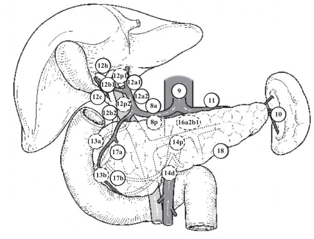
VỊ TRÍ CÁC NHÓM HẠCH
Nguồn: theo Miyazaki (2015) [1]
Nhóm hạch Định nghĩa
1 Nhóm hạch cạnh phải tâm vị
2 Nhóm hạch cạnh trái tâm vị
3 Nhóm hạch dọc theo bờ cong nhỏ dạ dày
4 Nhóm hạch dọc theo bờ cong lớn dạ dày
5 Nhóm hạch trên môn vị
PHỤ LỤC 2
XẾP LOẠI SỨC KHỎE THEO THEO HỘI GÂY MÊ HOA KỲ - ASA
(American Society of Anesthesiologist)
Sức khỏe tốt | |
ASA 2 | Có bệnh nhưng không ảnh hưởng đến sức khỏe và sinh hoạt hàng ngày. |
ASA 3 | Có bệnh ảnh hưởng đến sinh hoạt hàng ngày (đái đường có biến chứng mạch máu, bệnh phổi tắc nghẽn, cơn đau thắt ngực…). |
ASA 4 | Có bệnh nặng đe dọa tính mạng của BN (ung thư, suy tim, suy thận, phình động mạch chủ bụng, hen phế quản nặng…). |
ASA 5 | Tình trạng bệnh nhân rất nặng, hấp hối không có khả năng sống được 24 giờ dù có phẫu thuật hay không phẫu thuật. |
ASA 6 | Bệnh nhân chết não |
Có thể bạn quan tâm!
-

 Kết Quả Điều Trị Phẫu Thuật Nội Soi Hỗ Trợ Cắt Khối Tá Tụy.
 -
 Nghiên cứu ứng dụng phẫu thuật nội soi hỗ trợ cắt khối tá tràng đầu tụy điều trị u vùng bóng vater - 20
 -
 Nghiên cứu ứng dụng phẫu thuật nội soi hỗ trợ cắt khối tá tràng đầu tụy điều trị u vùng bóng vater - 21
 -
 Nghiên cứu ứng dụng phẫu thuật nội soi hỗ trợ cắt khối tá tràng đầu tụy điều trị u vùng bóng vater - 23
 -
 Nghiên cứu ứng dụng phẫu thuật nội soi hỗ trợ cắt khối tá tràng đầu tụy điều trị u vùng bóng vater - 24
 -
 Nghiên cứu ứng dụng phẫu thuật nội soi hỗ trợ cắt khối tá tràng đầu tụy điều trị u vùng bóng vater - 25
Xem toàn bộ 208 trang tài liệu này.
PHỤ LỤC 3
PHÂN LOẠI TNM THEO AJCC – 2010 (Nguồn: AJCC – 2010 [1])
UNG THƯ TÁ TRÀNG VÀ BÓNG VATER
T: Kích thước khối u
U nguyên phát không đánh giá được | |
T0 | Không có bằng chứng của u nguyên phát |
Tis | Ung thư tại chỗ |
T1 | U xâm lấn lớp niêm mạc và dưới niêm với kích thước u ≤ 1 cm (ung thư tá tràng); U khu trú tại cơ vòng Oddi (U Vater) |
T2 | U xâm lấn lớp cơ hoặc kích thước u ≥ 1 cm (ung thư tá tràng); u xâm lấn cơ tá tràng hoặc kích thước u ≥ 1 cm (u Vater) |
T3 | U xâm lấn tụy và lớp mỡ quanh tụy |
T4 | U xâm lấn mô mềm quanh tụy hay các cơ quan lân cận ngoài tụy |
N: Di căn hạch vùng
Chưa đánh giá được hạch | |
N0 | Chưa có hạch di căn vùng |
N1 | Hạch di căn vùng |
Hạch vùng bao gồm các nhóm: 13 (a, b), 17 (a, b), 14 (p, d), 12b, 8 (a, p) và *5, *6 (*không bắt buộc) | |
M: Di căn xa
Không thể đánh giá hạch di căn xa | |
M0 | Không di căn xa |
M1 | Di căn xa |
Di căn xa: di căn phổi, xương, màng phổi, gan, phúc mạc, não, tuyến thượng thận, không phải hạch vùng (nhóm 9, 10, nhóm 16), da… | |