- Reduce pain and discomfort: Daily monitor the level of pain and swelling of the parotid gland, testicles (in case of orchitis), epigastric pain (pancreatitis).
+ Pain relief using heat therapy.
Hot compresses are the best pain relievers. For patients with parotitis: Apply hot compresses to the jaw area. You can use 30 grams of crushed mung beans mixed with vinegar and apply to the swollen area or grind gac seeds, soak them in alcohol and rub on the swollen area. For patients with orchitis: Wear tight underwear to suspend the testicles. For patients with pancreatitis: Apply hot compresses to the epigastric area.
+ Rest helps relieve pain. Patients must lie still during fever and swollen glands, limit movement for the first 4-5 days.
+ Use pain relievers such as Paracetamol or Aspirin. Use anti-inflammatory Corticosteroids for patients with orchitis.
When using Aspirin and Corticoids for patients, it is necessary to give careful instructions to the patient when using it because it has many side effects, causing stomach irritation, stomach bleeding, so when giving it to the patient, it is recommended to take it with milk or during meals. Patients in pain often cannot sleep. Nurses should encourage and advise patients to sleep on time, avoid noise that affects the patient's sleep. Mild sedatives can be used for patients.
- Enhance nutrition for patients: Patients are often tired, have poor appetite due to pain and difficulty swallowing. Nurses need to guide patients in preparing and choosing food. Should eat liquid foods that are easy to swallow in the first few days. Eat many meals to ensure adequate protein and vitamins.
- Health education: As soon as a patient is admitted to the hospital, the patient and his/her relatives must be instructed on the ward rules in a gentle manner so that the patient can feel secure during treatment. The patient must be in an isolation room.
There should be a consultation about the disease and its progression so that the patient knows how to take care of himself and avoid spreading it to those around him. Know how to detect side effects of the drug. Instruct the patient on self-care: Dressing, resting, applying heat.
5.5. Evaluation
Review the patient care process to see if it meets the expected goals? It is considered good care if after one week the parotid gland gradually shrinks, the pain is reduced, and the symptoms of abdominal pain and difficulty swallowing gradually decrease and gradually disappear.
VALUATION
* Distinguish between true and false from sentences 1 to 6 by marking x in column A if the sentence is true and column B if the sentence is false:
STT
Question | A | B | |
1 | Mumps is a disease caused by a virus. | ||
2 | Mumps is transmitted through the digestive tract. | ||
3 | Mumps is common in children aged 5-9 years old. | ||
4 | Mumps only causes inflammation of the parotid gland, not inflammation of other glands. | ||
5 | Mumps viral parotitis never becomes suppurative. | ||
6 | Mumps orchitis patients will not have children. |
Maybe you are interested!
-
 Managing medical ethics education for nursing students in medical colleges - 32
 -
 Describe the causes of rickets.
 -
 Describe the clinical symptoms of acute appendicitis.
 -
 Transmission Methods Using Wavelength Division Multiplexing
 -
 Assessing customer satisfaction with the quality of medical examination and treatment at Ba Ria General Hospital - 11

* Choose the best answer for questions 7 to 10 by circling the first letter.
7. Early diagnosis of mumps when finding the following symptoms:
A. Sudden high fever, severe headache.
B. Found 3 Rillet and Barthez pain points
C. Painful swelling of the salivary glands
D. Increased salivation
8. Treatment of mumps is mainly:
A. Use antibiotics early
B. Symptomatic treatment
C. There is no specific medicine
D. Anti-inflammatory with Corticoids
9. The most important care for mumps patients is:
A. Fully comply with medication orders.
B. Closely monitor the patient's progress
C. Ensure rest and nutrition
D. All of A, B, C
10. To prevent mumps, it is necessary to do the following well:
A. Do not gather during an epidemic
B. Throat swab culture to find healthy carriers
C. Regular vaccination
D. Early isolation of sick people
Lesson 10
DIPHTHERIA PATIENT CARE
TARGET
1. Describe the causes and transmission of diphtheria.
2. Describe the clinical symptoms and complications of the disease.
3. Describe the patient care plan.
CONTENT
1. General
1.1. Definition
Diphtheria is an infectious, toxic disease caused by Coryne Bacterium Diphteriae. The first lesion is a pseudomembrane in the upper respiratory tract, from here bacterial toxins cause local and systemic damage.
1.2. Pathogens
- Caused by diphtheria bacteria (Coryne Bacterium Diphteriae).
- The bacteria are Gram (+) bacilli, shaped like drumsticks or dumbbells.
- Staining shows the image of white blood cells arranged in V, M,... with colors depending on the staining method (See books on microbiology)
- Bacteria secrete very toxic toxins.
For example, 1 mg of diphtheria toxin kills 1,000 guinea pigs, each weighing 250 g, within 96 hours.
- Resistance: Good, lives long in the patient's pseudomembrane and throat.
+ In low light conditions, bacteria can survive for months. In sunlight, bacteria die after a few hours.
+ On toys, clothes, etc., bacteria can live for several days.
+ Bacteria are destroyed at 58 o C within 10 minutes.
+ Phenol 1% solution, Sublime 1% kills bacteria in 1 minute
1.3. Epidemiology
- Source of infection:
+ Mainly sick people
+ Healthy people carry the virus
- Transmission route: Through the respiratory tract
+ Mainly direct transmission
+ Indirect transmission through utensils, toys, and clothes carrying white blood cells.
almost
- Sensory block: Most common in children from 1 to 9 years old. If vaccinated about
80% of children are protected.
2. Clinical symptoms
2.1. Ordinary pharyngeal diphtheria
2.1.1. Incubation period: 2 to 5 days.
2.1.2. Onset: The disease usually begins slowly with the following signs:
+ Mild fever 37 o 5C to 38 o
+ Tired, poor play, poor appetite.
+ Pale skin
+ Runny nose on one or both sides
+ Feel small, mobile, painless lymph nodes in the neck
+ Red throat, tonsils sometimes have faint white spots
- If detected at this stage: Swab the throat to find bacteria, treat promptly for high treatment results.
2.1.3. Full release:
- Toxic infection syndrome.
+ Mild fever 38 o C to 38 o 5C but pulse is quite fast, blood pressure is slightly low
+ Hard neck lymph nodes, mobile, painless
+ Tired, painful swallowing, pale.
+ Oliguria, urine has albumin
- White throat syndrome
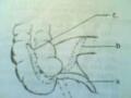
+ The white pseudomembrane is usually on one side of the tonsils or both sides, and can cover the uvula and soft palate.
+ Characteristics of pseudomembrane: Quite tough, sticks tightly to the throat, difficult to peel off, if peeled off it will bleed, put in water the pseudomembrane will not dissolve.
2.1.4. Progress
- Early treatment will improve the condition, and the pseudomembrane will fall off after 24 to 48 hours.
- Late treatment can cause diphtheria to spread to the larynx, causing laryngeal diphtheria or severe diphtheria, causing malignant diphtheria, with a high risk of death.
2.2. Laryngeal diphtheria
Usually occurring after pharyngeal diphtheria, the disease is common in children from 2 to 5 years old. The disease progresses through 3 stages:
+ Hoarseness and loss of voice stage: Lasts about 1 to 2 days, the child has a low fever of 38 degrees Celsius, fatigue, hoarse voice and a barking cough, then a hoarse cough, then loss of voice, unable to speak.
+ Dyspnea stage: Laryngeal dyspnea appears.
* Grading:
Grade 1: Intermittent dyspnea, increased by stimulation
Grade 2: Continuous difficulty breathing, struggling (best with tracheostomy) Grade 3: Rapid, shallow breathing, cyanosis, lethargy
+ Asphyxiation stage:
The child gradually becomes faint, motionless, comatose, turns purple and may die.
2.3. Malignant diphtheria
- Primary malignant diphtheria: Appears on the 1st or 2nd day of the disease
- Secondary malignant diphtheria: Appears on the 10th to 15th day of the disease.
* Clinical symptoms:
- Severe poisoning syndrome:
+ High fever, fatigue, pale skin, vomiting, painful swallowing
+ Dengue fever, nosebleeds, subcutaneous bleeding, internal bleeding.
+ The lymph nodes in the neck swell and stick together into a non-moving mass, causing the neck to bulge.
+ Low blood pressure, rapid pulse, muffled heart sounds, rapid heart rate, galloping rhythm, enlarged liver, little urination, urine with Albumin, high blood urea.
- Pseudomembrane: Rapidly spreading, gray in color, easy to bleed, bad breath
- Development
- Acute course after 24 to 36 hours
+ Rapid progression: Death after 5 to 6 days due to suffocation or hemorrhage
+ Subacute progression:
Initially, the patient had improvement on the 5th and 6th days, and on the 10th and 15th days, hematemesis and myocarditis appeared. The patient died suddenly due to irreversible circulatory collapse.
3. Complications
3.1. Cardiovascular complications: Common in diphtheria
- Cardiovascular disorders: Appear early in the first days of the disease. Usually the pulse is fast, if more severe, the pulse is fast and weak, blood pressure gradually drops leading to cardiovascular collapse, high risk of death.
- Myocarditis: can appear early in the first days, usually appears in the 2nd or 3rd week of the disease, very susceptible to acute heart failure and sudden death.
3.2. Neurological complications
Can appear early but usually in the 2nd or 3rd week of illness.
- Palatal palsy
- Paralysis
The above two signs usually recover after a few months.
3.3. Kidney complications
Glomerulonephritis or tubular nephritis. If severe acute renal failure, the risk of death is high for the patient.
core.
4. Treatment
Neutralize toxins as soon as possible with SAD:
+ Light: From 20,000 to 30,000 units
+ Severe: From 80,000 to 100,000 SAD units injected once subcutaneously or ½ dose intramuscularly when injected, it is necessary to test the reaction of serum diluted 1/1000 in physiological saline, 0.1ml injected intradermally, read the results after 20 minutes, positive reaction is: Red halo more than 10mm.
- Kill bacteria with antibiotics:
+ Penicillin G 50,000 to 100,000 IU/kg/day
+ Erythromycin:
Children: 40mg/kg/day Adults: 1.5g/day
- Supportive treatment: Vitamin C, cardiovascular support, sedatives, Corticoids for severe cases, tracheostomy.
5. Care
5.1. Assessment: Care assessment to establish baseline information about the patient's current condition.
- Ask about the disease in detail.
* Attention:
+ What day of illness?
+ Clinical symptoms from the first signs, pay attention to signs of mild fever but the patient is very tired, fussy, and plays poorly.
+ Is the epidemiological situation related to the patient but are there children around who have diphtheria and have they been fully vaccinated?
- Complete and thorough physical examination.
+ Regarding respiratory status: Count breathing rate, breathing pattern, determine laryngeal dyspnea.
Cyanosis, increased secretion, cough…
+ Circulatory status: Pulse count often shows rapid pulse. Blood pressure measurement often shows decreased blood pressure, listen to heart sounds.
+ Urinary condition: Oliguria, anuria
+ Nervous system: Fatigue, restlessness, agitation, paralysis...
+ Hoarseness, loss of voice
+ General condition: Temperature, pale skin, pseudomembrane development and color, bleeding...
+ Vital signs disorders due to infection and poisoning.
+ Airway obstruction due to increased secretion of phlegm, widespread pseudomembrane... Serious complications due to late detection, untimely treatment, poor care...
5.2. Care plan
Based on the data collected through the assessment, the following plan can be made:
- Basic care
- Monitor vital signs.
- Ensure air, prevent respiratory failure
- Monitor and prevent complications
- Take specimens for testing and monitor test results
- Monitor for signs of laryngeal diphtheria and malignant diphtheria.
5.3. Plan implementation
- Basic care
+ Resting the patient in an isolation room is very important, especially when there are complications of myocarditis. Absolute rest time is from 2 to 3 weeks or maybe from 40 to 50 days in a private, airy, bright, quiet room.
+ Diet: Eat enough nutrients, eat liquid foods in the acute phase, eat lots of fruit, drink lots of water. If the patient has pharyngeal paralysis, feed the patient with a thick consistency that is easier to swallow than liquid foods, and give them a little water at a time. If the patient cannot swallow, feed them through a tube.
+ Oral hygiene in the morning, evening and after meals. Personal hygiene to avoid ulcers.
- Monitor vital signs:
+ Count pulse, measure blood pressure every 30 minutes, every hour, every 3 hours
+ Measure temperature every 3 hours. When the patient has a high fever, the condition is severe and must be treated.
- Ensure ventilation:
+ When there are signs of increased secretion, wipe and suction the patient's sputum.
+ To reduce the patient's difficulty breathing, have the patient lie with the head elevated or use oxygen.
+ Prepare tools and medicine to help the doctor when a tracheostomy is indicated and care for the tracheostomy site like wound care.
- Track test results:
+ Take throat swab for testing
+ 24-hour urine measurement and urine test
+ Electrocardiogram
- Monitor to prevent complications: Especially cardiovascular complications. Let the patient rest completely, limit visitors to avoid noise.
- Health education: provides knowledge to:
+ Patients combine with the best doctor to achieve high efficiency in treatment
+ Diphtheria is dangerous but preventable: Vaccinate, do not gather children in crowded places where there is an epidemic. Isolate patients for at least 21 days. Patients must be discharged from the hospital after 3 negative throat swabs.
5.4. Assessment: Diphtheria patients present within 48 hours and have no complications. If treated well and treated well, the results will be good. If treated early (using SAD and antibiotics early), the risk of death is less than 5%. If treated late, from the 4th day onwards, the risk of death increases 20 times.
VALUATION
1. Could you please explain the definition, cause and pathogenesis of diphtheria?
2. Can you describe the clinical symptoms of diphtheria?
3. How do you present your assessment, diagnosis, and implementation of a care plan for diphtheria patients?
* Choose the one correct answer for the following sentences.
4. Fever in common diphtheria has the following characteristics.
A. Oscillation
B. Fever
C. High fever
D. Mild fever
5. Early signs of diphtheria and laryngitis are:
A. High fever
B. Shortness of breath
C. Discoloration of the pseudomembrane
D. Hoarseness
6. The incubation period of common pharyngeal diphtheria is:
A. 1 day to 2 days
B. 2 days to 5 days
C. 5 days to 10 days
D. 10 to 12 days.
7. What are the signs of malignant diphtheria?
A. Green skin
B. Runny nose on both sides
C. bulging neck
D. Mild fever.
Lesson 11
CARE FOR PATIENTS WITH WHOOPING COUGH
TARGET
1. Describe the epidemiological causes, clinical symptoms, treatment and prevention of whooping cough.
2. Plan care for a patient with whooping cough.
CONTENT
1. General
1.1. Definition
Whooping cough is an acute infectious disease caused by whooping cough bacilli, transmitted to patients through the respiratory tract. Clinical manifestations are characterized by particularly violent coughing attacks.
1.2. Pathogen: Bordetella-Gengou is an aerobic Gram (-) double-pointed bacteria cultured in Bosdel-Gengou medium and grows after 24 hours. The bacteria are not resistant to temperature: Under sunlight, the bacteria die after 1 hour, at 35 0 C, they die after 30 minutes.
1.3. Epidemiology
+ Source of disease: are patients with whooping cough. The disease spreads most quickly in the first week of the disease, with few symptoms of respiratory tract inflammation and the first coughing fits.
+ Transmission: whooping cough is transmitted through the respiratory tract by bacteria in droplets from the nose and mouth of a patient when coughing or sneezing, directly infecting healthy people.
+ Susceptibility: All ages, genders, ethnicities, and geographic regions can get whooping cough, but children aged 1-6 years are more susceptible; the younger the child, the more severe the illness.
+ Immunity: After getting sick, you have lifelong immunity.
2. Pathogenesis
Whooping cough bacilli enter the respiratory epithelium, multiply, and do not penetrate the blood vessels. There, they inhibit the activity of epithelial cells, causing acute inflammation of the respiratory tract and stimulating the mucosa to increase mucus secretion. Lesions occur mainly in the bronchi and bronchioles.
Bacterial toxins directly stimulate the nerve receptors of the respiratory mucosa, causing typical coughs, and on the other hand, affect the central nervous system. Here, the toxins directly affect the respiratory center in the medulla oblongata, causing respiratory disorders. If severe, it can lead to respiratory arrest. The toxins can also cause foci of excitement in the respiratory center, resulting in prolonged reflex coughs. The spread of toxins in the central nervous system can lead to encephalitis (a serious complication of whooping cough).
3. Symptoms
3.1. Clinical: (Typical Clinical Form)
3.1.1. Incubation period: 2 - 30 days (average 5-12 days) completely silent.
3.1.2. Onset period: (also known as the period of inflammation). Usually from 3-14 days with the following symptoms:
- Mild fever, gradually increasing.