- Patients are cared for both physically and mentally.
- No side effects of the drug.
- Vital signs and test results are fully monitored and recorded.
- Patients are instructed on rest, diet, self-exercise and massage, and comply with the doctor's treatment and care instructions.
VALUATION
Maybe you are interested!
-
 Describe the clinical symptoms of acute appendicitis.
 -
 Medical Service Projects, Health Care
 -
 Physical Health Care for Workers
 -
 Health care for the elderly in Duc Hue district, Long An province - 1
 -
 Criteria for Evaluating Public Investment Management for Health Care
1. Describe the definition of heart failure.
2. List some common causes of left heart failure.
3. Mark an x on the correct answer below:
A. High blood pressure causes right heart failure
B. Edema often appears in right heart failure.
C. When dealing with heart failure patients, be gentle and ask easy-to-answer questions.
D. Limit salt in the daily diet when caring for patients with renal failure.
heart
E. Electrocardiography is valuable in diagnosing heart failure.
4. Choose the best answer:
4.1. Causes other than right heart failure:
a. Mitral valve stenosis
b. Chronic lung diseases
c. Pulmonary infarction causing acute cor pulmonale
d. Scoliosis, thoracic deformity
e. Coarctation of the aorta
4.2. When assessing a patient with heart failure, the nurse needs to assess:
a. Ask about history of cardiovascular disease
b. History of medications used
c. Previously contracted diseases
d. Observe for difficulty breathing
e. All of the above
CARE OF PATIENTS WITH ACUTE PANCREATITIS
LEARNING OBJECTIVES
1. Describe the causes and pathogenesis of acute pancreatitis.
2. Describe the clinical symptoms, treatment principles and complications of acute pancreatitis.
3. Develop a care plan for patients with acute pancreatitis.
1. PATHOLOGY
1.1. Etiology
- Gallstones.
- Parasites: roundworms are the most common cause.
- Virus: mumps.
- Alcohol causes acute and chronic pancreatitis.
- After abdominal surgery, endoscopic retrograde cholangiography.
- Abdominal trauma.
- Malnutrition.
- Due to drugs, especially corticosteroids.
- Due to perforated gastric and duodenal ulcers adhering to the pancreas.
- Duodenal diverticulum.
- Pancreatic duct bifurcation.
1.2. Pathogenesis
- Duct theory: due to reflux of bile and duodenal juice into the pancreas, possibly due to stones stuck in the ampulla of Vater, spasm of the Oddi sphincter, or possibly due to increased pressure.
pressure in the biliary tract due to roundworm. This theory is not accepted because currently
Reflux can occur in normal individuals or during contrast cholangiography.
- Vascular theory: pancreatic infarction due to venous obstruction and the release of tissue kinases into the blood, which locally activate this enzyme.
- Hypersensitivity theory: also known as the X nerve theory because of the similarity in symptoms between parasympathetic hyperactivity and acute pancreatitis.
- Allergy theory: explains the phenomenon of disseminated vascular occlusion.
- Autolysis theory: explained on the basis of Trypsin activation by intestinal kinase reflux such as Enterokinase, bacterial leukocyte Kinase, lysosomal released by pancreatic lesions.
1.3. Clinical
- Pain: sudden, severe depending on the cause. May have different onsets.
- Vomiting: is a common symptom, occurring in about 70-80% of cases. Vomiting does not relieve pain.
- Abdominal distension: due to gastric and intestinal paralysis is also common. Some cases have signs of external abdomen, or signs of internal bleeding.
- Infection syndrome: depending on the cause, infection can come sooner or later.
- With hemorrhagic necrosis, the whole body shows signs of infection and severe poisoning.
- Jaundice: rare, if present is usually very severe.
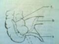
- Abdominal examination: Chauffard Rivet area is painful, Mallet Guy point is painful, Mayo Robson point is painful.
1.4. Testing
- Blood amylase: usually increases after about 4-12 hours of pain. With acute pancreatitis with edema, it will return to normal after about 34 days.
- Urinary amylase: increased slowly after 23 days.
- Blood lipase: often increases in parallel with blood amylase and is more specific. Exists in the blood for a long time.
- LHD and SGOT enzymes may increase in severe cases. These enzymes help assess prognosis.
- Blood calcium is often reduced in severe cases.
- PaO2 is often reduced.
- Blood formula: increased white blood cells, increased neutrophils, when white blood cells increase above 16,000/mm3, it has a serious prognosis.
- Ultrasound: large pancreas, poorer structure than normal.
- Unprepared abdominal X-ray: image of the guard bowel.
1.5. Diagnosis
1.5.1. Definitive diagnosis should be based on
- Acute abdominal pain in the epigastric region, left hypochondrium.
- Vomiting.
- Infection syndrome.
- Abdominal bloating.
- Painful pancreatic points.
- Ultrasound.
1.5.2. Differential diagnosis
- Perforation of hollow organs.
- Cholecystitis, acute cholecystitis.
- Acute intestinal obstruction and intussusception.
- Myocardial infarction: often occurs in the elderly with a history of angina, pancreatic examination is painless. Based on blood amylase.
1.6. Complications
- On site:
+ Pancreatic abscess
+ Pancreatic pseudocyst
+ Ascites: due to perforation or rupture of the pancreatic duct, pancreatic cyst ruptures into the abdominal cavity...
Whole body:
+ Collapsed lung, left lower lung inflammation.
+ Cardiovascular; digestive; kidney; metabolism.
1.7. Treatment
Help the pancreas rest
- Electrolyte replacement: in acute edematous pancreatitis, infuse about 23 liters/day of Ringer lactate solution and isotonic glucose.
Parenteral nutrition.
Pain relievers; atropine, Dolargan or Visceralgin
+ Atropine 1/4mg subcutaneous injection 12 mg divided 34 times a day.
+ Visceralgin tablets, 5ml tube.
Take 26 pills per day; intramuscular or intravenous injection 1/2 2 tubes per day.
Antibiotics:
+ In acute pancreatitis caused by alcohol, antibiotics are usually used slowly.
anti-infection
+ In acute pancreatitis due to bacteria, infection is very early so antibiotics should be used.
Early treatment usually involves using antibiotics against gram-negative bacteria such as
ampicillin, gentamycin. Ampicillin 500 mg intramuscular injection Gentamycin 80 mg intramuscular injection
+ In case of severe infection, it is necessary to combine 3rd generation Cephlosporin and 2nd generation Quinolone. In case of prolonged severe infection, it is necessary to use anti-anaerobic antibiotics; Imidazole, betalactamin, Macrolid (Clindacin, Dalacin).
- Treatment
pancreatitis
Ascaris: need to use
early use of anthelmintics;
Mebendazole (Fugacar) 100 mg tablets.
Treatment of acute pancreatitis due to stones: Oddi sphincter incision or lithotripsy.
1.8. Reserve
Regularly deworm, especially for people with a history of roundworms in the bile duct.
Good treatment for gallstones.
Limit alcohol.
Have a reasonable diet.
2. CARE FOR PATIENTS WITH PANCREATITIS
2.1. Comments
2.1.1. Diagnosis by medical history
See if there are any signs of infection?
Does the patient have abdominal pain? Where is the pain located and how is it severe?
Pain is intermittent or constant.
Pay attention to factors that increase pain
Does the pain increase when lying on your back and decrease when bending forward?
Does the patient have nausea or vomiting? Does vomiting relieve the pain?
Is there bloating?
- Does the patient have a history of alcohol consumption? Is there a history of acute pancreatitis due to worms or gallstones?
2.1.2. Observing the patient's condition
Infection status: dry lips, dirty tongue, tired, emaciated?
Mental status: restless, anxious, sweating or dizzy?
Observe the patient's pain-relieving posture.
2.1.3. Examination
Measure vital signs, paying attention to: temperature, pulse and breathing.
Examine the abdomen to determine where the pancreatic pain points are.
Review of paraclinical results:
+ Blood formula; high white blood cell count, increased neutrophil ratio.
+ High erythrocyte sedimentation rate.
+ Increased blood amylase or urine amylase.
+ Ultrasound and CT scan show images of pancreatitis.
2.1.4. Collecting data
Through medical records of treatment and care.
Through the patient's family.
2.2. Nursing diagnosis
Some possible major nursing diagnoses for patients with pancreatitis
grant:
Pain due to pancreatitis.
Vomiting due to stomach irritation
Abdominal distension due to gastroparesis
Increased body temperature due to infection.
Risk of pain shock.
2.3. Care planning
Rest and hygiene mode.
Diet.
Carry out the doctor's orders.
Monitor for possible complications.
Instruct patients on disease prevention.
2.4. Implement the care plan
2.4.1. Basic care
Let the patient rest completely in bed.
- Clean teeth, body, change clothes for patients daily, when patients vomit, must be taken care of cleanly and thoughtfully.
Measure pulse, temperature, blood pressure, electrocardiogram and check patient's consciousness.
- Place the nasogastric tube according to routine technique. Gently aspirate gastric fluid with a 50ml syringe, then connect the nasogastric tube to the drainage bottle.
Help the pancreas rest to reduce pain and secretion by fasting and gastric suction.
- Rehydration and electrolytes: patients are often dehydrated due to fasting, vomiting, and fever, so they need intravenous fluids.
- Oral feeding is only performed when pain symptoms have significantly reduced and the patient is fed from liquid to solid, starting with sugar water, then to porridge and porridge to reduce gastric secretion.
2.4.2. Follow your doctor's orders
- View medical records to carry out doctor's orders: IV fluids and other procedures.
- Take blood and urine for routine tests and mandatory tests for patients with acute pancreatitis such as: bleeding, urine, blood sugar, electrolytes (blood calcium), blood amylase...
- Gastric aspiration as indicated.
- Infusion: usually for acute pancreatitis with edema, about 2-3 liters/day is given.
- Painkillers should only be used when fasting and suctioning do not relieve pain. Dolargan can be used but morphine should not be used because it can cause oddi muscle spasm.
2.4.3. Monitoring and prevention of complications
Monitor vital signs every 3 hours.
Monitor the patient's abdomen: distention, pain, dullness to percussion.
Hang a level I nursing monitoring board at the bedside for seriously ill patients.
Prevent and monitor complications;
+ Pancreatic abscess: severe infection, high fever 3940o lasting more than a week, very painful pancreatic area, examination shows a raised mass, determined by ultrasound or CT scan.
+ Pancreatic cyst: the patient's pain and fever are reduced but do not return to normal.
usually. on weekdays
rank
23 pancreatic examination
have a block,
press tight,
Blood amylase is 23 times higher, ultrasound has empty Echo mass.
+ Ascites: due to perforation or rupture of pancreatic ducts or pancreatic pseudocysts into the abdominal cavity.
- Clearly record the date, time, name of the nurse and the patient's condition on the comprehensive care and monitoring form.
- Report to the treating physician the patient's condition and follow daily medical orders.
2.4.4. Patient education
- Instruct patients to follow doctor's instructions, fasting, urine retention... and administrative regulations of the treatment department.
- Instruct patients on appropriate diet when allowed to eat (avoid fat, alcohol, beer) and schedule a follow-up appointment after surgery to detect distant complications.
Regularly deworm, especially when there is a history of roundworms in the bile duct.
Good treatment for gallstones.
Limit alcohol intake.
2.5. Evaluation
A patient with acute pancreatitis is considered well cared for when:
The patient's pain is relieved, vomiting has stopped, and he can eat and drink by mouth.
Infections are reduced.
Tests returned to normal.
The orders were carried out completely and accurately.
No complications occurred