2.3. Testing
Gastroscopy shows defects and induration at the lesser curvature on multiple films.
Figure 74.3. Rigid shape at small curve - subsidence shape
Maybe you are interested!
-

 Basic electronic engineering - City College of Construction. HCM Part 1 - 1
 -
 Adjusting Loan Policies Reasonable To Encourage And Facilitate Meeting Students' Learning Needs
 -
 Study on microbiological characteristics of foot ulcers in diabetic patients at the Department of Endocrinology - Diabetes, Bach Mai Hospital in 2021 - 11
 -
 Basic Burn Rate Experimental Formulas
 -
 Follow-up Table of Patients Maintaining ARV Treatment After 12 Months
3. Complications
3.1. Wasting, leading to death.
3.2. Stomach perforation.
3.3. Gastric bleeding.
3.4. Pyloric stenosis.
3.5. Metastasis to adjacent organs.
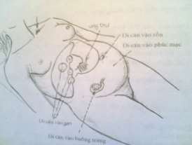
Figure 74.4. Metastasis of gastric cancer
4. Treatment
4.1. Need to detect at early stage
4.2. Explain and encourage the patient and family to transfer to a higher level facility early for diagnosis and treatment. Do not state clearly that this is cancer, causing the patient to become pessimistic and abandon treatment.
VALUATION
Please choose the best answer for the following questions: Question 1: Complications of stomach cancer:
A- Wasting leading to death. Gastric bleeding. Pyloric stenosis. Metastasis to neighboring organs. Gastric perforation.
B- Wasting leading to death. Gastritis. Pyloric stenosis. Metastasis to neighboring organs. Gastric perforation.
C- Wasting leading to death. Gastric bleeding. Cardiac stenosis. Metastasis to neighboring organs. Gastric perforation.
D- Wasting leading to death. Gastritis. Cardiac stenosis. Metastasis to neighboring organs. Gastric perforation.
Question 2 : Treatment of stomach cancer in primary health care:
A- Need to detect at early stage. Give painkillers, sedatives. B- Need to detect at early stage. Give antibiotics, vitamins. C- Need to detect at early stage. Use chemical treatment.
D- Need to detect at an early stage. Explain and encourage the patient and family to transfer to a higher level for treatment.
Question 3 : Paraclinical values in diagnosing stomach cancer:
A- Unprepared stomach X-ray in standing position shows a crescent of air under the diaphragm. Gastroscopy.
B- Unprepared gastric X-ray in standing position showing air-fluid level.
Gastroscopy
C- Unprepared gastroscopy in standing position with fluid in the abdomen. Gastroscopy. D- Gastroscopy showing defect and induration at the lesser curvature. Gastroscopy
Lesson 75
KIDNEY STONES
TARGET
1 Describe the causes of kidney stones
2. Describe the clinical symptoms of kidney stones
3. Describe the treatments for kidney stones.
CONTENT
1. General
Kidney stones are a common disease. It accounts for about 45% of all urinary tract stone diseases. Kidney stones are caused by many combined factors. It causes dangerous complications for patients.
2. Causes
Kidney stones are caused by many factors.
2.1. Local causes
- Urinary tract infection
- Damage to the subcellular region of the renal papilla (in case of infection or poisoning) forms a calcification and stones will form from that calcification.
- Urine retention for a long time will cause infection and stones.
2.2. Systemic causes
- Due to metabolic disorders of Oxalates, Urates, especially calcium metabolism disorders. Calcium increases beyond normal levels in urine. Increased calcium due to diet or endocrine disorders, especially due to hyperparathyroidism.
- Vitamin A deficiency: Creates conditions for keratinization of the renal intercellular tissue.
3. Pathology
3.1. Number of stones: Usually there is only one or more stones.
3.2. Location of gravel
- Stones located in the renal parenchyma: Usually small, fixed and underdeveloped.
- Stones located in the renal pelvis: More common, usually localized in the lower pelvis, fixed, only causing damage to one area of the kidney.
- Renal pelvis stones: The most common and dangerous, especially coral stones, occupy the entire renal pelvis and calyx, severely damaging kidney function.
3.3. Renal parenchyma
When the stones stay for a long time and gradually get bigger, they affect kidney function.
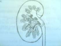
Figure 75.1. Renal pelvis and calyceal stones Figure 75.2. Hematuria or urinary obstruction during exercise
4. Symptoms
4.1. Functional symptoms
4.1.1. Typical renal colic
Colicky pain on one side of the lumbar region, radiating forward along the ureter and ending in the external genitalia. The pain often appears after heavy labor or long travel.
Along with the pain, the patient may have painful urination, frequent urination, or bloody urine. There may be nausea and vomiting. The pain will go away when the patient urinates out the stone or will be relieved by rest.
4.1.2. Hematuria : Common with the following characteristics:
- Blood in urine.
- Appears immediately after renal colic and gradually decreases when the patient rests.
- Often recurs after strenuous activity or a lot of walking.
4.1.3. Pyuria : Pyuria occurs when the kidney is severely infected. Patients with pyuria should consider kidney stones.
4.1.4 Passing stones in urine : rare, if passing stones in urine helps make the diagnosis more accurate.
4.2. Physical symptoms
4.2.1. Examination while the patient is in pain
- There is a reaction in the lumbar region.
- press the ureter and the patient's back pain points.
- Gently punching the patient's lower back is very painful.
4.2.2. If the kidney is dilated
- Examination shows signs of kidney touch and kidney floating.
- Examination in the early stages often does not show enlarged kidneys.
Figure 75.3. How to make the kidney touching and kidney floating signs
4.3. Subclinical symptoms
4.3.1. Urine : Depending on the level of kidney damage and infection, urine tests will show: Red blood cells, white blood cells, pus cells...
4.3.2. X-ray (upper line)
4.3.2.1. Unprepared kidney scan : Shows image of stones.
4.3.2.2. Intravenous pyelography (IV) : Shows whether the kidneys have stones or not and determines kidney function.
4.3.2.3. Retrograde pyelography (UPR) : Injecting photosensitive dye back from the ureter to the kidney, then taking a picture.
5. Complications
5.1.Pyelonephritis
The patient had high fever, pain in one side of the waist, pus in urine, blood in urine. Examination showed muscle reaction in the lumbar region.
The disease progresses in stages and often recurs.
5.2. Perirenal inflammation
The patient's whole body collapsed, with high and fluctuating fever and pain in the lumbar region. The lumbar region was swollen, red, and tender to the touch.
Must have surgery to drain pus.
5.3. Pyonephritis (pyonephritis)
This is a serious complication due to urine retention, infection and then kidney pus retention. The patient's whole body collapses, pus in the urine, and the kidney is enlarged and painful.
5.4. Anuria
This is a dangerous complication. Due to the stones from the renal pelvis moving down to the ureter and getting stuck, completely and suddenly blocking both ureters. Due to the kidney losing its ability to filter and excrete urine, it leads to anuria.
Early treatment is needed to avoid prolonged urinary retention in the kidneys, which can easily lead to kidney failure. Emergency kidney stone removal or drainage.
6. Treatment
6.1. Disease prevention
- Regular deworming is needed to avoid metabolic disorders.
- Ensure a balanced and reasonable diet, food should be varied.
- Give enough water to patients who have to lie down for a long time (spinal paralysis, spinal tuberculosis, bone fractures).
6.2. Internal treatment
- Applied in cases where the stones are small and can move out naturally or to prevent recurrence.
- Pay attention to your diet: eat lots of fruits, vegetables, and milk. Limit meat or foods high in calcium (depending on the type of stone).
- Use each course of eastern and western diuretics.
- Used in combination with vasoconstrictors such as Prostigmin or muscle relaxants such as Atropine or Nospa.
- Use antibiotics in cases of infection.
6.3. External treatment
Need to send patients to higher level for surgical treatment.
VALUATION
Please choose the best answer for the following questions: Question 1: Local causes of kidney stones:
A- Urinary tract infection. Damage to the supracellular region of the renal papilla. Long-term urinary retention.
B- Urinary tract infection. Damage to the subcellular region of the renal papilla. Long-term urinary retention.
C- Urinary tract infection. Damage to the supracellular region of the renal parenchyma. Long-term urinary retention.
D- Urinary tract infection. Damage to the subcellular region of the renal parenchyma. Long-term urinary retention.
Question 2 : Systemic causes of kidney stones:
A- Metabolic disorders of Oxalates, Urates... Especially Calcium.
Vitamin A deficiency.
B- Metabolic disorders of Oxalates, Urates... Especially Calcium.
Vitamin B deficiency.
C- Metabolic disorders of Oxalates, Urates... Especially Calcium.
Vitamin C deficiency.
D- Metabolic disorders of Oxalates, Urates... Especially Calcium.
Vitamin D deficiency.
Question 3 : Common locations of kidney stones:
A- Stones can be located in the renal pelvis, renal pelvis, and renal glomerulus.
B- Stones can be located in the renal calyx, renal parenchyma, or renal glomerulus. C- Stones can be located in the renal pelvis, renal parenchyma, or renal glomerulus. D- Stones can be located in the renal pelvis, renal parenchyma, or renal calyx.
Question 4 : Symptoms of pus in kidney stones:
A- rare, if found helps diagnose more accurately.
B- Appears immediately after renal colic and gradually decreases with rest.
C- When the kidney is severely infected. Patients with pus in urine should think of kidney stones. D- Often recurs after strenuous activity or a lot of walking.
Question 5: Physical symptoms of kidney stones during pain:
A- There is peritoneal reaction. Pressing the ureter and costovertebral points causes pain. Lightly punching the patient's lumbar region causes a lot of pain.
B- There is a muscle reaction in the lumbar region. Pressing the ureter and costovertebral points causes pain. Lightly punching the lumbar region causes a lot of pain.
C- Abdominal wall stiffness. Pressing the ureter and costovertebral points causes pain.
A light punch to the patient's lower back is very painful.
D- Stiffness in the lumbar region. Pressing the ureter and costovertebral points causes pain. Lightly punching the lumbar region causes a lot of pain.
Question 6 : Treatment of anuric complications of kidney stones:
A- Must place a bladder catheter to drain urine immediately. B- Drain urine through the skin and then surgically remove the stone.
C- Bladder catheter placement, renal pelvis drainage. D- Emergency surgery to remove stones or renal pelvis drainage.
Question 7 : Things to note in kidney stone prevention for patients who have to stay in bed for a long time: A- Patients should be allowed to move early and gently in bed.
B- Regular deworming is needed to avoid metabolic disorders. C- Give enough water.
D- Provide adequate nutrition and avoid stone-forming substances.
Question 8 : Applying medical treatment to patients with kidney stones:
A- Small stones can move out naturally, or to prevent stones from recurring.
release
B- Stones located in the renal pelvis or renal pelvis.
C- Stones located in the renal pelvis or in the renal parenchyma. D- Kidney stones without complications.
Lesson 76
URETERAL STONES
TARGET:
1. Describe the causes and pathology of ureteral stones.
2. Describe the symptoms of ureteral stones
3. Describe the treatment of patients with ureteral stones at the primary health care level.
CONTENT:
1. General
- Ureteral stones are a delayed surgical emergency.
- In some cases, stones on both sides of the ureter causing obstruction require emergency surgery. If not intervened promptly, the patient may die due to anuria.
- Most ureteral stones fall from the kidney 80%.
- There are some cases where small stones pass down to the bladder and the patient urinates out. The stones often stop at the narrow part of the ureter: the renal pelvis - ureter, the ureter crossing the iliac artery, the ureter close to the bladder.
2. Causes of disease
2.1. Primary stones
Usually 80% of kidney stones fall out.
2.2. Secondary stones
- Due to the consequences of some diseases such as: tuberculosis, syphilis, ureteral damage due to surgery causing ureteral stricture.
- Due to ureteral malformation: dilated ureter, ureter behind the vena cava, double ureter. Urine stagnates above the narrow area, sediments and forms stones.
3. Pathology
3.1. Pebbles
- Location of the pebble:
+ 70 to 75% of stones are in the lower third of the ureter.
+ 20 to 30% of stones are in the middle third and upper third of the ureter.
- Shape: Usually oval, like a peanut or rough like a strawberry.
- Pebble color:
+ Calcium oxalate stones are solid, shiny black.
+ Ivory white calcium phosphate stones.
- Size and number of stones: Usually the diameter of the stones is less than 1cm, in some cases the stones are as big as chicken eggs. Usually there is only one stone, in some cases there are two stones or in some cases the stones are arranged in a chain.
3.2. Ureteral stones
- In the ureter, there is often acute damage, edema of the mucosa, then fibrosis reaction leading to ureteral narrowing just below the stone.
- The ureter above the stone is dilated and the renal pelvis is also dilated, causing hydronephrosis, renal pus retention, and gradual destruction of kidney tissue.
4. Symptoms
4.1. Clinical symptoms
- Functional symptoms
+ Renal colic: Usually appears after exertion with the characteristics of lumbar pain, pain radiating to the front of the right or left hypochondrium, spreading down to the groin, ending at the external genitalia. Pain causes sweating. The pain can last for many hours. In many cases, severe pain can cause shock.
+ When there is urine retention in the kidney or ureter, the patient has dull pain and tension in the lumbar region.
+ When in pain, the patient may have vomiting and bloating.
+ Transient hematuria.
+ Painful and frequent urination when stones near the bladder irritate.
- Physical symptoms:
+ During the pain of ureteral stones, the patient's examination shows stiffness of the lumbar muscles, stiffness of the abdomen on the side of the ureter with stones, and abdominal distension.
+ When stones block the ureter, causing hydronephrosis and pyonephrosis, the kidney is found to be enlarged.
- Systemic symptoms:
+ Physical condition changes little when there is only one stone on one side.
+ The patient has a fever when the stone affects the ureter and has a urinary tract infection.
+ Bilateral ureteral stones cause urinary obstruction, causing the patient to collapse quickly because of high blood urea, oliguria, and anuria.
4.2. Subclinical symptoms
- Ultrasound shows image of stones, condition of renal pelvis, ureter above stones.
- Unprepared urinary tract imaging shows radiopaque ureteral stones.
- Contrast urinary system imaging shows the location of stones, the path of the ureter, and assesses kidney function.
- Retrograde ureterography and pyelography detect stones, especially non-obstructive stones.
light
5. Complications
- Urinary tract infection.
- Enlarged kidney with hydronephrosis and pus retention.
- Ineffective when there are stones on both sides causing urinary obstruction.
- Kidney failure.
- High blood pressure.
6. Treatment at primary health care facilities:
Same as kidney stone treatment.
6.1. Disease prevention
- Regular deworming is needed to avoid metabolic disorders.
- Ensure a nutritious and reasonable diet, food should be varied.
- Give enough water to patients who have to lie down for a long time (spinal paralysis, spinal tuberculosis, bone fractures).
6.2. Internal treatment
- Applied in cases where the stones are small and can move out naturally or to prevent recurrence.
- Pay attention to your diet: eat lots of fruits, vegetables, and milk. Limit meat or foods high in calcium (depending on the type of stone).
- Use each course of eastern and western diuretics.
- Used in combination with vasoconstrictors such as Prostigmin or muscle relaxants such as Atropine or Nospa.
- Use antibiotics in cases of infection.