1.2. Check the abdomen
Use observation and palpation methods.
Observe the belly area: stand behind and observe the size and uniformity of both sides of the belly. Normally, when a buffalo is full, the left belly is slightly larger than the right belly.
1.2.1. Observe the abdominal area
Volume of distended abdomen:
Maybe you are interested!
-

 Internal control of revenue and expenditure activities at Vietnam Academy of Traditional Medicine - 15
 -
 Request for Completion of Internal Control of Revenue and Expenditure Activities at Vietnam Academy of Traditional Medicine
 -
 Common Internal Auditing Issues
 -
 Internal control of revenue and expenditure activities at the National Children's Hospital - 2
 -
 The Nature and Role of the Internal Control System
+ Food retention in the stomach for a long time: overeating
+ Gas accumulation due to fermented food: bloating (buffalo and cow's abdomen is swollen on the side)
left)
chemical
+ Liver disease, kidney disease, bladder obstruction
+ Pregnant animals (buffaloes and cows have swollen belly on the right side) The belly volume shrinks:
+ Long-term hunger.
+ Prolonged diarrhea, prolonged pneumonia, gastrointestinal parasites
1.2.2. Checking the ruminant stomach
The ruminant stomach consists of 4 pouches: rumen, reticulum, omasum and abomasum. In the anterior stomach: rumen, reticulum, omasum do not have digestive glands but thanks to the rumen microorganism system, the fibers are fermented, converting cellulose into volatile fatty acids (acetic acid, propionic acid, butyric acid). These acids provide energy for ruminants. In addition, dead microorganisms will be decomposed by digestive juices and will be a source of protein for the body to be absorbed in the posterior stomach (abomasum).
Because of the abundant microflora, when livestock are sick, this microflora easily changes in quantity, affecting the health of livestock.
Rumen
Location: left side, front of the back thigh. Observing the hip socket:
+ Bloating, swollen rumen, loss of left flank.
+ Animals that starve for a long time will have deep sunken hips.
+ Rumen hyperphagia: enlarged lower left rumen.
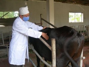
Figure 4.1: Palpation of the rumen of a cow
Yes, bee.
Location at the end of ribs 6 - 7 - 8 behind the heart, above the sternum. How to check: let the animal go down the slope, or use a stick to lift the animal up.
honeycomb stomach area. The animal will have a pain reaction due to the organs in the abdominal cavity pressing on the honeycomb stomach.
Yes, book leaves.
Location: on the right side between ribs 7 and 9 on a line parallel to the ground, from the shoulder blade joint.
How to check: use a 15cm long needle to poke into the position of the vellus. If the needle moves, the vellus is still moving. If the vellus is paralyzed, the needle will stand still. If the vellus is paralyzed, the animal will have a high fever.
Star fruit
Location: right hand, behind the stomach is a book
How to check: Right abdomen is large: dilated abomasum, inflammation due to parasites. Pressing on the animal will cause pain.
1.2.3. Simple stomach test
Big left abdomen: animal is overfed, bloated. Treatment is to induce vomiting and wash the stomach.
1.2.4. Checking stomach contents
By placing a probe in the abdomen of the buffalo to check the contents of the buffalo's stomach.
1.2.5. Bowel check
Rectal examination:
Purpose: pregnancy check (buffaloes), bladder check, bowel check.
Organ location: 15 cm deep on the left is the rumen, on the right is the uterus, above is the kidney, below is the bladder.
Visual inspection of stool:
Amount of stool: In case of diarrhea, the animal will have loose, watery stools in the early stages. If the animal has a high fever, the stools will be concentrated, constipated, and scanty.
Treatment of constipation: use vaseline, glycerol or rectal water to soften stool.
Stool color:
White feces in piglets and calves: indigestion in young cattle, paratyphoid in pigs.
Bright red stool: colitis, rectal bleeding, disease
dysentery..
Black and red stools: due to bleeding in the upper intestines such as the stomach.
thick, duodenum
Green stool: due to food, infectious diseases, digestive juices in stool, porcine paratyphoid, diarrhea, etc.
Yellow stool: due to duodenal juice, intestinal mucosa, in diseases caused by
E.coli , swine fever, coccidiosis,..
Stool with a lot of mucus: chronic constipation, mucous enteritis
Pseudomembranous stool: intestinal mucosa peels off, a sign of enteritis
heavy.
State of division
Stool with lots of gas bubbles: enteritis caused by food contaminated with microorganisms
ferment.
Parasites in feces
1.2.6. Liver Test
Location of the liver: The liver is a large mass, located in front of the abdominal cavity. In ruminants, the liver is located on the right side of the abdominal cavity.
Pig liver has 4 basic lobes: front, back, right and left. In cattle, the liver is not lobed but is a large block of about 5kg.
Liver function: secrete bile, accumulate glycogen, fat-soluble vitamins, protect, decompose old red blood cells, create blood... If the disease occurs in the liver, it often reduces liver function, often causing some pathological conditions such as:
Enlarged, pale liver, enlarged gallbladder (paratyphoid in pigs) Necrosis due to parasites.
Sclerosis
Liver and gallbladder diseases often have jaundice symptoms.
2. Common internal diseases of the digestive system
2.1. Stomatitis
2.1.1. Causes
Primary cause:
Due to the oral mucosa being stimulated by mechanical impacts (hard food, teething, etc., stimulating the oral mucosa → causing inflammation
Due to thermal stimulation (food, drinks that are too hot...) Due to chemical effects
Secondary causes
Due to inflammation from other organs
Consequences of systemic diseases (deficiency of C, A, anemia)
Due to infectious diseases: foot and mouth disease, cattle cholera, swine fever, etc.
2.1.2. Symptoms
Acute form:
Drooling, dry nasal mucosa, red or patchy red, slow eating, difficulty chewing.
The tongue is gray-white, if the disease is severe, the tongue is swollen and painful. Chronic form
Similar to the acute form but prolonged, cattle eat poorly and become increasingly thinner, the oral mucosa thickens, becomes concave, not smooth, the tongue surface is ulcerated, and the mucosa inside the cheeks is inflamed and ulcerated.
2.1.3. Prognosis
In the primary form, the animal will recover after about 7-10 days. If not treated with care, the disease will last longer and the animal will become emaciated.
2.1.4. Diagnosis
Based on symptoms of the disease
2.1.5. Treatment
Good nursing Use of medication
Use antiseptic solution to wash mouth area. Apply antibiotic to the ulcer area.
Supplement vitamins A, CB 2 , PP
2.2. Sore throat
2.2.1. Causes
Primary cause:
Due to weather changes, resistance is reduced.
Due to mechanical impact: objects in food, mucosal abrasion or use of esophageal catheter.
Due to irritation of the throat mucosa by toxic chemicals. Secondary cause
Secondary to infectious diseases: flu, tuberculosis, septicemia,....
Due to inflammation from other areas: stomatitis, rhinitis, laryngitis,...
2.2.2. Symptoms
Acute pharyngitis the animal shows pain, reduces appetite, stretches head and neck, front legs scratch the ground, and pretends to chew.
Drooling. Mouth may be inflamed, tongue coated with plaque, bad breath, occasional vomiting.
The nasal discharge is clear at first, then becomes cloudy like pus, mixed with food particles.
eat.
Cough, wet cough, painful, if inflammation spreads quickly to the larynx, cough becomes more severe. Palpate the throat area of the animal, pain, discomfort and cough, if pseudomembranous inflammation
and honeycomb inflammation, the inflamed area is very hot, the lymph nodes under the jaw are swollen.
Blood test: increased white blood cell count, increased neutrophils, decreased eosinophils and lymphocytes.
Urine test: acid urine, Albuminuria.
2.2.3.Prognosis
Acute catarrhal pharyngitis usually resolves after 1-2 weeks. If the disease is pseudomembranous or ulcerative, the disease will last longer, and if pus-causing bacteria invade, it will turn into purulent pharyngitis. Pharyngitis can progress to catarrhal pneumonia, pneumonia caused by foreign bodies entering the lungs, laryngeal edema, and severe disease can cause sepsis.
2.2.4. Diagnosis
It is necessary to understand the characteristics of the disease. You can open the animal's mouth to examine the throat, and see the swollen and red mucosa.
2.2.5. Treatment
Nursing, good care. Medication.
Use oil to reduce inflammation. Use antiseptic solution to rinse throat.
If there is a high fever, use antibiotics. If there is suffocation: perform a tracheostomy. If there is purulent inflammation: clean the pus.
Support, support.
2.3. Esophageal obstruction
2.3.1. Causes
Because livestock quickly swallow root foods or dry powdered foods without giving them water.
Due to swallowing foreign objects.
Due to anesthesia while the esophagus still contains food. Due to secondary causes of esophageal diseases.
Due to poisoning.
2.3.2. Symptoms
Choking, neck always stretched high to swallow, worried posture, shaking head, mouth watering with vomiting reaction.
When the esophagus is blocked → air cannot escape → flatulence. If a foreign object compresses the trachea → the animal has difficulty breathing, suffocation. Palpate the swollen esophagus.
2.3.3. Prognosis
If the blockage is caused by soft objects, the stool may pass into the stomach and resolve on its own within a few hours to a day.
If the blockage is caused by large, solid objects, the disease will last for a long time → the animal cannot eat, the esophagus may tear, bloating → suffocation and death.
2.3.4. Diagnosis
Diagnosis is based on symptoms.
2.3.5. Treatment
Nursing: keep the animal in a high head, low body position, give it plenty of water to drink. Intervention:
If the foreign object is stuck in the back of the throat: use a tool to remove the foreign object.
If the foreign object is in the neck: if the foreign object is soft (massage, give water to drink), if the foreign object is hard, iron, smooth (use paraffin or vegetable oil to inject into the esophagus and then gently stroke it out of the mouth with your hand).
If the foreign object is stuck in the lower part: use an esophageal catheter to slowly push it in. Use esophageal stimulants.
If there is flatulence: treat flatulence with trocar. If esophageal obstruction is caused by a sharp foreign object: surgically remove the foreign object.
2.4. Crop disease in poultry
2.4.1. Causes
Due to eating indigestible foods, fermented foods. Due to being poisoned by strong chemicals.
Secondary to stomatitis, crop paralysis or vitamin deficiency. In pigeons, it can also be caused by milk that has accumulated in the crop and fermented, causing inflammation.
Due to parasites that live in the crop.
2.4.2. Symptoms
The animal is weak, has poor appetite, drinks more water than usual, often stretches its neck and makes swallowing movements. The crop is swollen, full of air, and when pressed, the animal feels pain. The animal often burps or has a runny, sour-smelling discharge.
When the animal is turned upside down, water flows out of the beak. The water is cloudy gray and has a sour, putrid smell. The animal often develops diarrhea.
2.4.3. Treatment
Nursing; fast for a few days, tilt head upside down, stroke food from crop to beak to expel all food.
Intervention
Instill antiseptic substances into the crop: 2% boric acid, 1% iron sulfate, 1% alum, 1% sodium bicarbonate.
In case of disease caused by parasites, bleach must be used.
Cut open the crop, remove all food, wash with 1% potassium permanganate solution, then sew.
again.
2.5. Rumen paralysis
2.5.1. Causes
Improper livestock care and feeding methods, buffaloes and cows eat a lot of concentrated feed, little green feed; eat too monotonous food or change food too suddenly. When food is scarce, cattle are hungry, eat moldy, rotten straw, lack vitamins, causing weakness. Cattle may have heart, liver, kidney diseases, metabolic disorders, or other chronic diseases. Strongly stimulating food makes rumen peristalsis too excited, in the later stages it will reduce muscle tone, rumen peristalsis decreases and then paralysis.
In addition, the management of livestock is not reasonable, livestock are overworked, and grazing conditions change. Due to the secondary development of other diseases such as internal medicine (overeating rumen, bloating rumen, honeycomb gastroenteritis caused by foreign objects, peritonitis, peritoneum); infectious diseases (flu, septicemia); parasitic diseases (liver flukes, blood parasites) or acute poisoning. Pathological effects hinder the activity of the central nervous system, autonomic nervous system, then hinder the activity of the pregastric, causing the rumen muscles to reduce peristalsis and leading to paralysis.
2.5.2. Symptoms
Cattle eat less, prefer roughage to concentrate, thirsty, rumination is reduced or stopped, rumen motility is poor or absent; belching, slightly foul smelling. The animal likes to lie down, tired, dry oral mucosa. Palpation of the rumen through the rectum shows food like thick porridge, left abdomen is swollen, difficulty breathing. Loose stools mixed with mucus, when enteritis develops, loose stools are foul smelling. If the disease is severe, the animal has convulsions and dies.
2.5.3. Lesions
The disease causes the volume of the rumen and abomasum to increase, the rumen area to sink, the food in the abomasum dries up, the rumen is filled with foul-smelling mucus, and the stomach lining is inflamed or hemorrhagic.