- Deep layer: flexor digitorum profundus, flexor pollicis longus, pronator quadratus muscle.
3.2. Posterior forearm region: the muscles of the posterior forearm region are arranged in 2 layers:
- Shallow layer: includes two groups:
+ Outer group: brachioradialis muscle, extensor carpi radialis longus, extensor carpi radialis brevis.
Maybe you are interested!
-
 Evaluation of the effectiveness of selective tibial nerve resection in the treatment of lower limb muscle spasticity sequelae - 19
 -
 Anatomy and physiology of the digestive system - Master. Doctor. Tran Quang Thao - 10
 -
 For Investment in Construction of Headquarters, Factories, Canteens, Rest Houses (Not Including Land Use Rights Value).
 -
 Anatomy and physiology of the digestive system - Master. Doctor. Tran Quang Thao - 7
 -
 Clinical features, endoscopic images of laryngoscopy and pathological anatomy of vocal cord cysts - 7
+ Posterior group: extensor digitorum, extensor carpi radialis, elbow muscles.
- Deep layer: abductor pollicis longus, extensor pollicis brevis, extensor pollicis longus, extensor pollicis indexis, supinator.
The nerve that innervates the muscles of the posterior arm is the radial nerve, which is responsible for supinating the hand and extending the fingers and hand.
4. Muscles in the hand
- The hand is limited from the furthest wrist crease to the tips of the fingers, divided into two parts: the palm and the back of the hand.
- The muscles of the hand include the dorsal interosseous muscle, the palmar interosseous muscle, and the worm muscle. These muscles are innervated by the median and ulnar nerves.
B. LOWER LIMBS
I. LOWER LIMB BONES
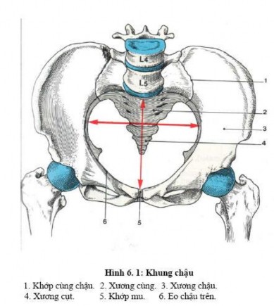
1. Pelvis
1.1. Description
The pelvis is a pair of fan-shaped bones, the pelvis on one side is continuous with the pelvis on the opposite side and the sacrum behind to form the pelvis. The pelvis is shaped like a basin, narrowed in the middle, the constriction is the superior iliac isthmus. The pelvis has the function of containing the abdominal organs and transferring the weight of the body to the lower limbs.
1.2. Structure
Embryologically, the pelvis is composed of three bones joined together. The center of the connection is the acetabulum, which contains vestiges of Y-shaped cartilage.
- Iliac bone: above, consists of two parts: the body and the wings of the iliac bone.
- Pubic bone: in front, consists of: body and two branches: superior and inferior.
- Ischial bone: posterior, consists of ischial body and ischial ramus.
1.3. Anatomical characteristics
The pelvis is a flat bone with 2 surfaces and 4 borders.
1.3.1. Outer surface: in the middle there is a cup-shaped depression called the acetabulum for the femoral head to articulate. Above the acetabulum is the gluteal surface for the gluteal muscles to attach. Below the acetabulum is the obturator foramen, covered by an obturator membrane. In front of the obturator foramen there is a groove (tube) for the obturator blood vessels and nerves to pass through.
1.3.2. Inner surface: in the middle is the arc, running diagonally from top to bottom to front; The two arcs of the two pelvic bones and the posterior sacral protuberance form the superior iliac isthmus. The superior iliac isthmus divides the pelvis into two parts, the upper part is the greater pelvis, the lower part is the lesser pelvis. The superior iliac isthmus is very important in obstetrics. Above the arc is the pelvic fossa, behind the pelvic fossa there is an ear-shaped articular surface called the tympanic surface to articulate with the sacrum. Below the arc is the square surface corresponding to the posterior acetabulum, below the square surface is the obturator foramen.
1.3.3. Upper border: is the iliac crest, the highest point of the iliac crest is at the level of the 4th lumbar vertebra.
1.3.4. Lower border: formed by the fusion of the ischial ramus with the lower ramus of the pubic bone.
1.3.5. Front bank: has the following details:
- The anterior superior iliac spine is an important anatomical landmark.
- Pubic mound.
- The pubic tubercle is attached to the inguinal ligament. The inner and lower surfaces of the pubic tubercle have a pubic surface to articulate with the pubic bone on the opposite side.
1.3.6. Posterior edge: also has many concave and convex areas, with details:
- Posterior superior iliac spine.
- Large sitting defect.
- Sit down.
- Small sitting defect.
- Sitting position: is the place that bears the entire body weight when sitting.
2. Femur
The femur is a long bone consisting of a shaft and two ends.
2.1. Bone shaft
The triangular prism has three faces: front, inside, outside; three edges: inside, outside and back. The back edge is convex and sharp, called the rough line, with many muscle attachments.
2.2. Top end
There are femoral head, femoral neck, greater trochanter and lesser trochanter.
- Femoral head: 2/3 spherical shape, pointing up, in and out.
- Femoral neck: connects the head with the two trochanters, tilts upwards and inwards. The axis of the neck meets the axis of the body at an angle of 130 0 called the tilt angle, helping the femur to move easily.
- Greater trochanter: is the attachment site of the rotator cuff muscle, can be felt and located in living people.
- Lesser trochanter: on the back and inside of the femur.
2.3. Lower end
The lower end has the medial and lateral condyles. The outer surface of the lateral condyle has the lateral epicondyle process; the inner surface of the medial epicondyle has the lateral epicondyle process and the adductor tubercle.

3. Patella
It is a flat triangular bone, the base is above the apex and the bottom is below. The patella is encased in the quadriceps tendon and is therefore called a sesamoid bone. It plays a role in the extension of the lower leg.
4. Tibia
The tibia is the main bone of the lower leg, bearing almost the entire weight of the body from above. The tibia is a long bone with a shaft and two ends.
4.1. Bone shaft
A slightly convex triangular prism. It has three faces and three edges:
- Of the three surfaces, the inner surface is flat and close to the skin.
- Of the three borders, the anterior border is sharp and close to the skin. The anterior border as well as the inner surface are close to the skin, so when the tibia is broken, it can easily pierce the skin, causing an open fracture. At the same time, the bone is difficult to heal when damaged.
4.2. Top end
Widely flared to support the femur, including:
- Medial convexity.
- Lateral condyle, more convex than medial condyle, below and behind has fibular articular surface to articulate with the upper end of the fibula.
The upper surface of each condyle has a corresponding superior articular surface for articulation with the femoral condyle.
The anterior aspect of the two condyles has a tubercle just under the skin called the tibial tuberosity, where the patellar ligament attaches.
4.3. Lower end
Smaller than the top, including:
- Medial malleolus: formed by the lower part of the inner part of the head, can be felt under the skin.
- Lower articular surface: articulates with the upper surface of the talar trochlea.
- Fibular defect: on the outer surface of the joint of the lower end of the fibula.
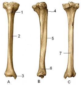
Tibia Shape
A. Front view B. Outside view C. Back view
1. Tibial tubercle 2. Medial surface 3. Medial malleolus 4. Superior end
5. Body 6. Lower end 7. Posterior surface
5. Fibula
The fibula is a long, thin bone that lies on the outside of the tibia.
5.1.Bone body
The shaft of the bone has three surfaces and three borders.
5.2. Top end
Also called fibular head, articulation with tibial-fibular joint surface, palpable under the skin.
5.3. Lower end
Flatter and more pointed than the upper end, forming the lateral malleolus, the lower end of the lateral malleolus being lower than the lower end of the medial malleolus. The lower end of the fibula and the lower end of the tibia form the tibiofibular arm which plays a very important role in walking.
6. The bones of the foot
The foot bones include: the ankle bones, the metatarsals, and the toe bones.
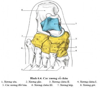
6.1. The ankle bones
Consists of 7 bones arranged in two rows:
- Posterior row: has two bones, the talus and the calcaneus.
- Front row: has 5 bones: navicular bone, cuboid bone and three cuneiform bones.
6.2. Metatarsal bones
There are 5 metacarpal bones from inside to outside: Metatarsal I,... Metatarsal V. Each bone has a base, body and head.
6.3. The metatarsal bones
Each toe has three bones: the proximal phalanx, the middle phalanx, and the distal phalanx. The exception is the first toe, which has only two bones: the proximal phalanx and the distal phalanx. Each bone also has three parts: the phalanx base, the phalanx body, and the phalanx tip.
II. LOWER LIMBS
1. Buttock muscles
The gluteal region is an area with many important blood vessels and nerves passing from the pelvis to the lower limbs. The gluteal muscles are divided into two groups with different functions.
- The iliotibial trochanter muscle group includes the following muscles: tensor fascia femoris, gluteus maximus, gluteus medius, gluteus minimus, and piriformis. These are the muscles that extend and rotate the thigh.
- The ischial tuberosity pubis muscle type includes the following muscles: obturator internus, gastrocnemius, quadratus femoris, and obturator lateral. These muscles mainly rotate the thigh externally.
2. Thigh muscles
The thigh is bounded above by the inguinal crease anteriorly and the gluteal crease posteriorly. Below by a horizontal line three finger widths above the base of the patella. The thigh muscles are divided into two regions.
2.1. Anterior thigh muscles: include two muscle groups.
- The anterior muscle area is the thigh flexor and leg extensor area including the quadriceps, sartorius and iliopsoas muscles, mainly innervated by the femoral nerve.
Action: stretch the leg, the rectus femoris muscle also helps to flex the thigh.
- The inner muscle area is the thigh adductor region, including the pectoral muscle and 3 adductor muscles: adductor longus, adductor brevis and adductor magnus, whose function is to close the thigh, controlled by the obturator nerve.
.
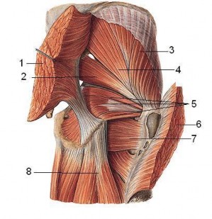
Figure of the muscles of the buttocks
1 and 6. Gluteus maximus 2. Piriformis 3. Gluteus medius
4. Gluteus minor 5. Obturator internus and gastrocnemius 7. Quadriceps femoris
2.2. The muscles of the posterior thigh: include three ischial tuberosity muscles: semimembranosus, semitendinous and biceps femoris, which are responsible for extending the thigh and flexing the leg. The nerves supplying the muscles of the posterior thigh are branches of the sciatic nerve.
Popliteal fossa
Is a 4-sided diamond-shaped pit located behind the knee joint containing the vascular and nerve bundles of the region.
skillful
Four sides are
- Above and inside are the semitendinous and semimembranous muscles.
- On the outside is the biceps femoris muscle.
- The two lower edges are the two heads of the gastrocnemius muscle.
In the popliteal fossa there are the tibial nerve, the popliteal artery, the popliteal vein, some other blood vessels, nerves and superficial lymph nodes of the popliteal region, in which there are two special superficial nerves: the medial calf cutaneous nerve separated from the tibial nerve and the lateral calf cutaneous nerve separated from the common peroneal nerve; The special superficial veins include the small saphenous vein that goes from the dorsal venous arch up to the popliteal region and then goes deep to empty into the popliteal vein, the small saphenous vein is the vein that is often affected by varicose veins.
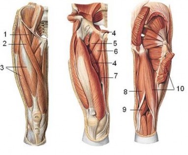
Figure 10. 5. Thigh muscles
1. Iliopsoas muscle 2. Segura muscle 3. Quadriceps muscle 4. Adductor longus muscle
5. Plectrum muscle 6. Adductor brevis muscle 7. Adductor magnus muscle 8. Semitendinosus muscle
9. Semimembranosus muscle 10. Biceps femoris muscle
3. Muscles of the calf area
The lower leg is limited above by a line passing under the tibial tubercle, and below by a line passing through the two malleolus. The muscles of the lower leg are divided into