- Number of tumors: ……….. 1 mass 2 - 3 masses > 3 masses
- Tumor size 1:.................mm < 5cm 5 – 10 cm > 10cm
- Tumor size 2:.................mm < 5cm 5 – 10 cm > 10cm
- Tumor size 3:.................mm < 5cm 5 – 10 cm > 10cm
- Location: Stomach Duodenum Small intestine Colon Rectum
Other locations................................................................................................
- Portal vein thrombosis: No Yes Not described
- Abdominal lymph nodes: No Yes Not described
- Other abnormalities on abdominal ultrasound ...................................................................
D5. Bowel flow imaging: Do not do
Can you see the tumor ? Can you see the tumor?
D6. CT scan: Do not do
Can you see the tumor ? Can you see the tumor?
- CT scan conclusion:
- Ascites: None Little Moderate Much
- Size: clearly stated..................mm < 5 cm 5 – 10 cm > 10cm
- Quantity: clearly state the quantity ……….. 1 block 2 - 3 blocks > 3 blocks
- Location: Stomach Duodenum Small intestine Colon Rectum
Other locations: ...................................................................................................
- Properties on CT: Increase density Decrease Copper Mixture
Necrosis............................... Drug absorption................................
- Portal vein thrombosis: No Yes
- Abdominal lymph nodes: No Yes
- Other abnormalities:………………..........................................................................
D7. MRI scan: Do not do
Can you see the tumor ? Can you see the tumor?
- MRI scan conclusion:
- Ascites: None Little Moderate Much
- Size: clearly stated..................mm < 5 cm 5 – 10 cm > 10cm
- Quantity: clearly state the quantity ……….. 1 block 2 - 3 blocks > 3 blocks
- Location: Stomach Duodenum Small intestine Colon Rectum
Other locations: ................................................................................................................
- Properties: Increased signal Decreased Same Mixed
- Portal vein thrombosis: No Yes
- Abdominal lymph nodes: No Yes
- Other abnormalities:………………..........................................................................
D8. PET-CT: No Tumor detected Tumor detected Description: …………………………………………………………………………….......
D9. Biopsy / CDHA : No Yes Result: …..……………………………...
D10. Other surveys (specify):……………………………………………………………….
E. DIAGNOSIS
E1. Preoperative diagnosis: ........................................................................................................
E2. Postoperative diagnosis: .......................................................................................................…………..
F. SURGICAL METHODS Session Emergency
PTV :………………………..
F1. Translation : No little medium much Not described
F2. Peritoneal metastasis : Yes No Not described F3. Liver metastasis: Yes No Not described F4. Metastasis elsewhere: Yes .................................. No
F5. Abdominal lymph nodes: Yes No Not described
F6. Tumor location and corresponding tumor size:
Esophagus: Upper 1/3...................... Middle 1/3................................ Lower 1/3................................
Stomach: Cardia............. Antrum............. Fundus................. Pylorus ................
BCL................... BCN...................................
Duodenum: D ........... .....................
Jejunum ..................... Ileum ...........................
Colon: Cecum.......... Ascending colon............ Transverse colon............ Descending colon............
Singma phone............... Right phone................ Left phone................
Rectum .......... Anus .......... Other locations :..........................................................
F7. Color: Pinkish white / yellowish white Pinkish brown, gray
F8. Type of surgery:
Laparoscopic Surgery Open Surgery Laparoscopic Conversion to Open Endoscopic Assistance
Biopsy exploration Open or bypass (cannot remove tumor)
Wedge resection Local tumor resection (through the anus or rectum)
Cut the OH with tumor: Immediately repeat digestion Take it out for HMNT. Amputate the TT
1 position 2 positions 3 positions 4 positions 5 positions
Specifically: Esophagus Stomach Duodenum Jejunum Ileum Cecum
Upward phone Horizontal phone Downward phone Singma phone Right phone Left phone
Rectum Anus Other: ............................................................................
F9. Lymph node dissection: Yes No
F10. Other: …………........................................................................................................
................................................................................................................................................................ G. PATHOLOGY: Number: ……………… ICD-10: ………….. ICD-0: …… GROSS IMAGE
G1. Number of tumors: (specify the number of tumors) ………………………………………………....
Classification of tumor number 1 tumor 2 – 3 tumors > 3 tumors
G2. Location and corresponding tumor size (mm):
Esophagus: Upper 1/3...................... Middle 1/3................................ Lower.................................
Stomach: Cardia............. Fundus............. Antrum............. Pylorus .................
BCL................... BCN...................................
Duodenum: D(1-4) ........... .....................
Jejunum ..................... Ileum ...........................
Colon: Ascending colon........... Transverse colon............ Descending colon............ Singma colon..............
Right phone................ Left phone................
Rectum ............... Anus .................... Location
other:.........................................
G3. Tumor density: Firm Soft Mixed Not described
G4. Tumor shape: Sphere, mass, polyp, lobule Flat, plaque, infiltrating Not described G5. Capsule, border: Yes No/Diffuse infiltration Not described G6. Tumor color: Pinkish white / yellowish white Pinkish reddish brown, gray Not described MICROSCOPE RESULTS
G7. Definitive diagnosis (cytological diagnosis)
Lymphoma Leiomyoma Rhabdomyosarcoma GIST Granuloma Lipoma
Hemangioma Melanoma Schwannoma Kaposi sarcoma Glomus tumor
Suspect: …………………………………………………………………………………...
IMMUNOHYTOCHEMISTRY
G8. Immunohistochemistry: To be done Not required If positive, required, but not yet done Marker Type: ......................................................................................................................
Result: ............................................................................................................................
G9. Suspected differential diagnosis: .....................................................................................
H. POSTOPERATIVE FOLLOW-UP
H1 . General results: Death, severe Good discharged from hospital
H2. Reoperation No Yes: (due to: …………………………)
H3 . Early complications: No Yes
Surgical site infection Pleural effusion Residual abscess Peritonitis
Urinary tract infection Anastomotic leak Pneumonia Postoperative bleeding
Multiple organ failure Urinary retention
H4. Long-term follow-up (assessment at the end of the study)
Died Date: ......../......../.............
Living with disease recurrence
Live without disease
Recording method: Telephone interview. Date of call:...................../Meeting:..................... Postoperative adjuvant treatment: No Yes
Chemotherapy Radiotherapy Chemoradiotherapy Targeted therapy Immunotherapy Other: …………………………………………………………………………….....
Survival time after surgery: …………(months)
Appendix 2
MICROPHOTOGRAPHY AND IMMUNOCHYTOCHEMISTRY
07 TYPES OF NON-EPHELIAL CARCINOMA
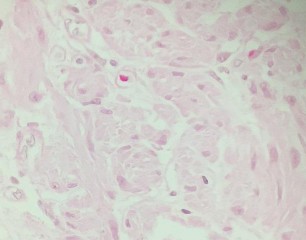
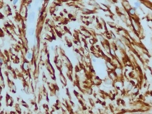
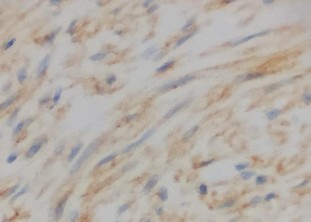
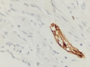
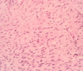
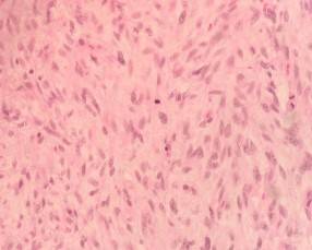
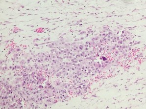
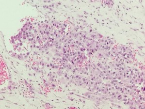
1. Malignant rhabdomyosarcoma (Patient: Pham Van T.)
| |
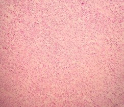
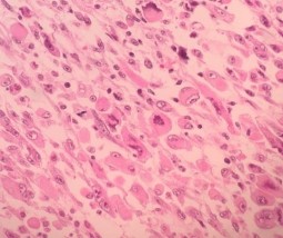
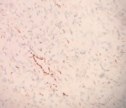
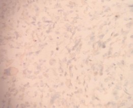
HE x 100: Spindle-shaped tumor cells, arranged bundle | HE x 400: Cytoplasm of tumor cells is pink, large nucleus, clear nucleolus, many mitotic divisions |
|
|
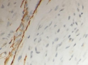
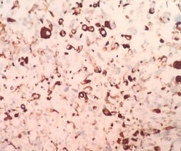
DESMIN: Cytoplasmic positive tumor cells | MYOGLOBIN: Granular positive with tumor cell cytoplasm |
|
|
SMA: Negative for tumor cells | DOG 1: Negative for tumor cells |

Maybe you are interested!
-
 Software Technology - Pham Hung Phu, Nguyen Van Tham Compiled - 36
 -
 Scientific commentary on the criminal code Volume 4 - Dinh Van Que - 2
 -
 Network design - Master Tran Van Long, Master Tran Dinh Tung Compiled - 24
 -
 Financial statement analysis of VT Van Xuan Joint Stock Company - 1
 -
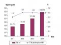 Divergence of Land Use Criteria in the Planning of Van Yen District in 2014 and Forecast for 2015 (Unit: %)
2. Liposarcoma (Patient: Pham Trong C.)
| |
HE x 100: Highly differentiated tumor component with tumor cells have wide vacuoles, nuclei at the edge of the cytoplasm | HE x 100: Anaplastic tumor component with spindle-shaped tumor cells and oval nuclei |
|
|
HE x 100: Anaplastic tumor component with spindle-shaped tumor cells, oval nuclei | HE x 400: Rich in mitotic figures |
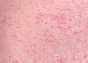
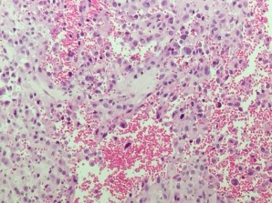
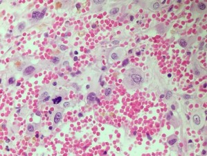
3. Angiosarcoma (Patient: Nguyen Van M.)
| |
HE x 100: Epithelioid tumor cells, interspersed with blood vessels | HE x 100: Epithelioid tumor cells, interspersed with blood vessels |
|
|
HE x 100: Epithelioid tumor cells, interspersed with blood vessels | HE x 400: Clear nucleus, distorted nuclear membrane, rich in division |
|
|
HE x 400: Clear nucleus, distorted nuclear membrane rich, multiplied | HE x 400: Clear nucleus, distorted nuclear membrane, rich in division |

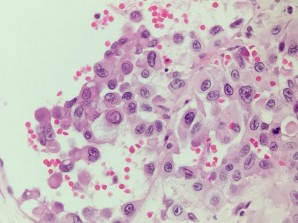
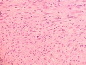
4. Gastrointestinal stromal tumor (GIST) (Patient: Nguyen Thi L.)
| |
HE x 100: Spindle cell tumor with oval nucleus, bundle | HE x 400: Spindle-shaped tumor cells, oval nucleus arrange into bundles |
|
|
DOG1: Strongly positive for nucleus and cytoplasm | CD117: Cytoplasmic positive |
|
|
S100: Negative for tumor cells, positive calculated with internal evidence. | SMA: Negative for tumor cells, positive calculated with internal evidence. |