According to the BRAF mutation test (50%), the first regimen uses Vemurafenib, when there is no longer response, the second regimen is used with Ipilimumab. In addition, recent research shows that 15.6% have KIT mutations in skin lesions. In these cases, Imatinib is the treatment of choice for patients [116]. Interferon-β, INF-2 or IFN-2b [126] are used as supportive and palliative treatment in advanced cases or used with chemotherapy to help control tumor progression and recurrence. [119].
1.3.9. Vascular glomus
Surgical treatment is the mainstay, ensuring a wide enough excision area to be safe and rarely reoccurring, but it is also necessary to provide periodic monitoring instructions to the patient. No supportive treatment is required [134, 138, 139].
1.3.10. Malignant granuloma
Mainly by surgery. For small tumors that can be removed through flexible endoscopy, radical surgery is performed when a malignant tumor is identified, invading the deep layer of the OTH wall [145]. The prognosis for recurrence with benign tumors is 2-8%; With malignant tumors, it is difficult to completely treat, the time for recurrence and metastasis is short (2 years) and death is usually within 3 years [141, 143, 168]. Radiation therapy and chemotherapy are not necessary for benign tumors and are ineffective for malignant lesions. Recently, we have seen reports of response when used with Pazopanib from several groups of authors such as Conley (2014), Lei Wei (2015) and Sachi Morita (2015) [169, 170].
1.3.11. Malignant rhabdomyosarcoma
With malignant rhabdomyosarcoma at OTH, radical surgery that removes the intestinal segment with the tumor is best. Multi-chemotherapy is applied including chemotherapy and radiotherapy (for tumors in the anorectal area or with regional metastases). resides in or behind the peritoneum, does not affect large blood vessels in the abdomen) [155, 171].
1.4. STATUS OF RESEARCH ON TYPES OF NON-Epithelial Gastrointestinal Duct CANCER IN VIETNAM
Since the 1970s, author Nguyen Duc Ninh described a case of a patient with a tumor in the duodenum with a tumor 1.5 cm in diameter, soft in density, and round in shape, causing severe gastrointestinal bleeding [172].
The article about the small intestine by author Nguyen Phuc Cuong and Nguyen Trung Tuan researched the pathological images of 38 small bowel tumors in the 10 years 1974-1983 in Viet Duc [173].
In 1986, the group of authors Nguyen Nhu Bang, Truong Nam Chi, and Pham Kim Binh in a pathological study of 422 cases of stomach cancer in 5 years (1976-1980) found that 4% were non-epithelial cancer, of which 9 were non-epithelial cancers. case of malignant lymphoma and 6 cases of malignant glioma [174].
In 1993, author Do Duc Van in his research on the surgical treatment of stomach cancer at the Viet-German hospital (1970-1992), commented that through 1908 people who received surgical treatment, there were 14 cases of malignant lymphoma and 8 cases of malignant lymphoma. case of malignant glioma [175].
In 1994, authors Doan Huu Nghi and Pham Hoang Anh through research on stomach cancer in Hanoi people from 1988 to 1992 summarized 1068 patients, including 1 case of sarcoma and 3 cases of malignant Schwann cell tumors. , however, the authors excluded from the study all gastric tumors whose tumor nature was unclear. Most likely, there were many stromal tumors among them, requiring immunohistochemistry to determine. This study is only at the statistical level and does not mention this type of tumor [176].
In 1995, a report at the 3rd national conference on radiology and nuclear medicine on adult small bowel intussusception: causes, diagnosis and treatment in 2 cases by Trinh Hong Son and Nguyen Duy Hue [ 84].
In 1995, through 359 cases of colon tumors operated on in Viet Duc in 8 years (1986-1993), there were 15 cases of non-epithelial tumors including 13 cases of malignant lymphoma and 2 cases of malignant lipoma [177].
In 1996, the article "VFM combined with severe gastrointestinal bleeding due to neuroma in the small intestine" by Trinh Hong Son and Cao Doc Lap raised the issue of difficulty in initial diagnosis and serious complications due to intestinal tumors. caused by immaturity [178].
In 1997, Trinh Hong Son researched "clinical forms of small bowel tumors" through 42 cases including benign and malignant tumors (1998). Silent or acute progression (gastrointestinal bleeding, VFM, intestinal obstruction, intussusception...). Clinical manifestations are diverse, most of which cannot be diagnosed before surgery. The main treatment is surgical resection of the small intestine [179].
In 1998, non-adenoepithelial gastric cancer was also researched and published in the Journal of Practical Medicine in 1998 on 23 cases in the period 1993-1999 at Viet Duc Hospital by authors Trinh Hong Son, Nguyen. Phuc Cuong, Do Duc Van. The majority of cases were admitted to the hospital because of abdominal pain, 1 case of tumor perforation and 2 cases of gastrointestinal bleeding, and 12 cases of abdominal tumors. Of these, 3 cases were malignant leiomyomas [180].
In 2000, another study on malignant leiomyomas of the small intestine by authors Trinh Hong Son, Pham Duy Hien and colleagues showed that through 12 cases of malignant leiomyomas of the small intestine operated on at Viet Duc Hospital in the early stages. 1990-1999, malignant leiomyomas accounted for about 16% of malignant tumors of the small intestine, most could not be diagnosed before surgery, most occurred in the jejunum, common clinical signs were abdominal pain, confusion. digestion and weight loss, especially physical signs of a palpable tumor in the abdomen and are common in men [181].
In 2000, in the period 1990-1999, with the article "Malignant lymphoma of the small intestine" by author Trinh Hong Son and his colleagues, within 10 years, only 13 cases of surgery were recorded at Viet Duc hospital [ 54].
In 2002, Le Dinh Roanh and colleagues in a study on histopathological classification of gastric cancer on 452 cases (2002) showed that gastric stromal tumors and malignant lymphomas accounted for an equally low proportion (1, 55%). The author used immunohistochemistry to diagnose difficult cases and discovered 1 case with c-kit mutation [182].
In 2002, author Nguyen Ngoc Hung studied 62 cases of non-epithelial tumors of the stomach operated on at Viet Duc hospital since 2002.
1995 to 2002 showed that the most common tumor was lymphoma (33.87%), followed by glioma (30.65%), leiomyoma (20.97%), and undifferentiated sarcoma. stroma: 9.68%), tumors of other components such as fibers, fat, and vessels are very rare. Malignant tumors are more common than benign; In the lymphoma group, men are more common than women; In the group of benign connective tissue tumors, women are more common than men; the common age of the whole group as well as each pathological type is 50-70 [15].
In 2002, a case of severe gastrointestinal bleeding due to a leiomyoma was reported by Trinh Hong Son and colleagues. A 43-year-old male patient was admitted to the hospital because of red blood in his stool. The tumor in the lead II wall probably protruded into the duodenal lumen. 3cm diameter, pathology result was malignant leiomyoma. At the same time, review domestic and foreign medical literature on this disease [183].
In 2005, statistics of VFM cases due to pathological small bowel perforation in 5 years in Viet Duc (2000-2004) by the group of authors Trinh Hong Son and colleagues, there were 2 cases due to malignant tumors: leiomyoma and tumor. lymphoma in a total of 14 patients [184].
In 2006, in the newspaper "Practical Medicine", the article "diagnosis and treatment of small intestinal stromal tumor" by authors Trinh Hong Son and Pham Gia Anh reported a case of small intestinal stromal tumor: male patient 41 years old. years old, had 2 surgeries to remove the tumor, received supportive treatment, then treated with the drug Glivec provided by Novartis. After 3 months, the abdominal tumor disappeared, the liver mass became smaller and there was no more fluid in the abdomen. This is the first patient in Vietnam to receive this drug [11].
In 2007, author Nguyen Van Mao's thesis researched histopathology and immunohistochemistry through 32 cases of malignant gastrointestinal stromal tumors and made recommendations for pathological diagnosis [64].
In 2011, author Nguyen Thanh Khiem's inpatient thesis was on "clinical and paraclinical characteristics and results of surgical treatment of primary gastrointestinal lymphoma at Viet Duc hospital" [14]
In 2011, author Bui Trung Nghia's inpatient thesis was on "Evaluation of clinical and paraclinical characteristics and results of surgical treatment of gastrointestinal stromal tumor (GIST) at Viet Duc hospital from January 1/2011." 2005 to December 2010” [12].
In 2012, author Trinh Hong Son clinically reported a case of a large rectal leiomyoma that the pathology easily confused between GIST and leiomyoma. Thanks to HMMD, the diagnosis was confirmed as leiomyoma. benign of the rectum [185].
In 2017, author Do Hung Kien with a study on the clinical and paraclinical characteristics of gastrointestinal stromal tumors (GISTs) at a stage where surgery to remove the tumor is no longer indicated, with CD 117 (+) and the results. Results of treatment of this group of 188 patients with imatinib and some related factors [13].
Chapter 2
RESEARCH SUBJECTS AND METHODS
2.1. RESEARCH SUBJECTS
These are patients diagnosed with non-epithelial cancer of the digestive tract who were operated on at Viet Duc University Hospital within 10 years, from January 2009 to April 2019.
2.1.1. Patient selection criteria
Each patient selected for the study met all of the following criteria:
- Patients regardless of age or gender.
- The patient is treated surgically.
- Pathology results confirmed the diagnosis of non-epithelial malignant tumors of the entire digestive tract including the esophagus, stomach, duodenum, small intestine, colon, rectum and anal canal .
- Have complete medical records with clinical symptoms and paraclinical results, surgical records, and pathology results.
2.1.2. Exclusion criteria
- The pathology result was OTH carcinoma.
- Pathology results were benign.
- Patients who do not have complete medical records or pathology results.
2.2. RESEARCH METHODS
2.2.1. Research design
Retrospective descriptive research method, patients were retrospectively reviewed on medical records of clinical and paraclinical symptoms, pathology, diagnostic imaging, and surgical procedures according to a systematic research medical record form. best. Fellows directly access data, analyze it, contact post-operative follow-up and evaluate results.
2.2.2. Sample size
Convenient sampling: All patients meeting research criteria had surgery within a 10-year period from January 2009 to April 2019.
2.2.3. Research time and location
- Time: from January 2009 to April 2019
- Research location: Viet Duc Friendship Hospital
2.2.4. Data collection method
Step 1: Get all results as non-epithelial tumors of OTH at the Department of Pathology: Including 2 sources
- Source 1: Get data from handwritten archives:
+ Get pathology results in original books at the pathology department from January 2009 to January 8, 2011: Read more than 40,000 pathology results in 8 notebooks from book number 52 to number 59.
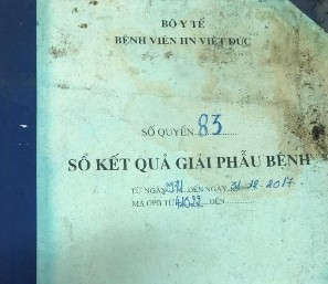
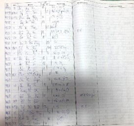
Figure 2.1. Pathology results book (January 2009 - January 2011)
+ In these more than 40,000 results, the diagnoses of retroperitoneal tumors, pelvic tumors and mesenteric tumors are eliminated to get the diagnoses of malignant non-epithelial tumors and benign ones (in indistinguishable cases). clearly understand the nature of the tumor and need immunohistochemistry to differentiate and confirm the diagnosis) of the esophagus, stomach, duodenum, small intestine, colon, rectum and anus.
- Source 2: Get data from the results storage software of the Pathology Department from January 8, 2011 to April 2019:
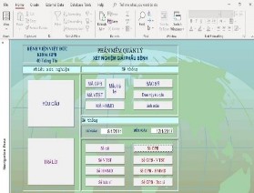
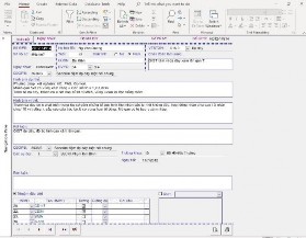
Figure 2.2. Pathology results management software (2011 – 2019)
Find each morphology code of the International Classification of Diseases for Oncology (ICD-O) of non-epithelial tumors
Table 2.1. Morphological code according to the International Classification of Diseases for Oncology
No
Vietnamese name | English name | ICD-O code | |
1 | Vascular glomus | Glomus tumor | 8711 |
2 | Granuloma | Granular cell tumor | 9580. 8620. 8622. 8600. |
3 | Malignant leiomyoma | Leiomyosarcoma | 8890. 8896 |
4 | Malignant rhabdomyosarcoma | Rhabdomyosarcoma | 8900. 8901. 8910. |
5 | Malignant lipoma | Liposarcoma | 8850 |
6 | Kaposi's cancer | Kaposi sarcoma | 9140 |
7 | Malignant melanoma | Melanoma | 8720. 8700. |
8 | Malignant angioma | Angiosarcoma Haemangioma | 9120 9150 |
9 | Malignant schwannoma | Shwannoma malignant | 9560 |
10 | Gastrointestinal stromal tumor | GIST | 8936. 8801 |
11 | Malignant lymphoma | Lymphoma malignant | 9591. 9590. 9650. 9675. 9680. 9684. 9687. 9705. 9970. |
Maybe you are interested!
-
 Study on clinical features, pathology and treatment results of non-epithelial gastrointestinal cancer at Viet Duc Hospital - 17
 -
 Research on current status and improvement of cropping system on coastal land of Thanh Hoa province - 19
 -
 Review of Research Status Related to the Topic
 -
 Hoang Thanh Phuc (2009), "Research on Current Status and Solutions for Developing Scattered Forestry Planting in Thai Nguyen Province", Master's Thesis,
 -
 Research on the current status and proposed solutions for conservation of Pseudotsuga brevifolia WC Cheng & LKFu, 1975 in Nguyen Binh district, Cao Bang province - 10