Table 3.11 Prevalence of autonomic dysfunction in Parkinson's disease
Survey test
Abnormality rate (n = 82) | |
Parasympathetic function | Frequency (%) |
Heart rate variability with deep breathing | 41 (50) |
Postural heart rate variability | 16 (19.5) |
Valsalva maneuver | 24 (29.3) |
Sympathetic function | Frequency (%) |
Isometric exercise – | 49 (59.8) |
Postural blood pressure variation | 5 (6.1) |
Recording of sympathetic skin response | 25 (30.5) |
Maybe you are interested!
-
 Techniques for Performing Autonomic Nerve Function Tests
 -
 Measure 4: Make Tools and Toys to Use in Children's Play Activities
 -
 Perspectives on Improving the Quality of Law Application in Resolving Land Use Rights Disputes at the People's Court
 -
 Internal control of revenue and expenditure activities at the National Children's Hospital - 2
 -
 “Cronbach's Alpha Cost Factor Scale”
The prevalence of autonomic dysfunction varied according to the tests. The highest rate of abnormalities in the Parkinson group was in the isometric exercise blood pressure variation test (59.8%).
Parkinson's disease
13
(15.9%)
44
(53.7%)
17
(20.7%)
8
(9.8%)

Normal
Sympathetic Parasympathetic
Sympathetic and parasympathetic
Figure 3.4 Distribution of autonomic dysfunction in Parkinson's disease
In 82 Parkinson's patients, when examined on 6 tests, only 13 cases were normal (accounting for 15.9%), the remaining 69 cases had at least 1 abnormal test.
survey, of which 44 cases (53.7%) had abnormalities affecting both the sympathetic and parasympathetic systems.
3.2.2 Characteristics of autonomic nervous system disorders in multiple system atrophy Table 3.12 Results of tests to assess cardiovascular autonomic nervous system function
on multiple system atrophy
Survey test
Average index (n = 45) | |
Parasympathetic function | Average ± Standard |
Deep breathing – BTNT (beats/minute) | 5.05 ± 3.27 |
Change your posture – Index 30:15 | 1.06 ± 0.08 |
Valsalva Test – Valsalva Index | 1.19 ± 0.15 |
Sympathetic function | Average ± Standard |
Isometric exercise – Diastolic BP (mmHg) | 4.36 ± 6.19 |
Postural blood pressure - Systolic BP (mmHg) | -18.64 ± 19.54 |
Ewing Total Score | 3.22 ± 0.96 |
BTNT: heart rate variability, BTHA: blood pressure variability
During inspiration and expiration, heart rate changes were small with a mean variation of 5.05 ± 3.27 beats/min. After standing, there was an increase in heart rate when standing compared to lying down, with a mean 30:15 ratio of 1.06 ± 0.08. The mean Valsalva index was 1.19 ± 0.15, indicating an increase in heart rate when blowing into a resistance tube.
After holding the blood pressure monitor for 3 minutes, there was no significant change in diastolic blood pressure with a mean blood pressure variation of 4.36 ± 6.19. After standing for 3 minutes, there was postural hypotension with a mean systolic blood pressure variation of -18.64
± 19.54.
The mean Ewing score for all five tests was 3.22 ± 0.96.
Table 3.13 Prevalence of autonomic dysfunction in multiple system atrophy
Survey test
Abnormality rate (n = 45) | |
Parasympathetic function | Frequency (%) |
Heart rate variability with deep breathing | 40 (88.9) |
Postural heart rate variability | 12 (26.7) |
Valsalva maneuver | 20 (44.4) |
Sympathetic function | Frequency (%) |
Isometric exercise – | 35 (77.8) |
Postural blood pressure variation | 15 (33.3) |
Recording of sympathetic skin response | 23 (51.1) |
The prevalence of autonomic neuropathy varies among tests.
The highest rate of abnormalities in the MSA group was in the heart rate variability test with deep breathing (88.9%). This was followed by abnormalities in the isometric exercise test (77.8%).
During electrical stimulation, 23 cases of MSA recorded no sympathetic skin response waves, accounting for 51.1%.
Table 3.14 Comparison of test results between MSA-P and MSA-C
Survey test
MSA-P (n = 18) | MSA-C (n = 27) | p | |
Deep breathing – BTNT (beats/minute) | 4.79 ± 3.31 (median: 3.5) | 5.23 ± 3.31 (median: 5) | 0.536 ** |
Change your posture - Index 30:15 | 1.04 ± 0.07 (median: 1.03) | 1.08 ± 0.09 (median: 1.06) | 0.093 ** |
Valsalva maneuver – Valsalva index | 1.16 ± 0.14 | 1.21 ± 0.17 | 0.340 * |
Isometric exercise – Diastolic BP (mmHg) | 4.78 ± 6.28 | 4.07 ± 6.25 | 0.714 * |
Postural blood pressure - Systolic BP (mmHg) | -18.06 ± 20.26 | -19.04 ± 19.43 | 0.871 * |
Ewing score | 3.39 ± 0.96 | 3.11 ± 0.96 | 0.349 * |
* t-test ** Mann-Whitney test BTNT: heart rate variability, BTHA: blood pressure variability
Compare 2 subgroups:
There were no differences in autonomic dysfunction indices between the MSA-P and MSA-C subgroups when examining each test separately (p > 0.05).
When examining the total Ewing score, there was also no difference in the degree of autonomic dysfunction between the MSA-P and MSA-C subgroups (p = 0.42).
Table 3.15 Comparison of autonomic dysfunction rates between Parkinson's and cerebellar multiple system atrophy
Survey test
MSA-P (n = 18) | MSA-C (n = 27) | p | Check determine | |
Frequency (%) | Frequency (%) | |||
Heart rate variability with deep breathing | 16 (88.9) | 24 (88.9) | 1,000 | Fisher |
Postural heart rate variability | 7 (38.9) | 5 (18.5) | 0.175 | Fisher |
Valsalva maneuver | 10 (55.6) | 10 (37) | 0.221 | χ 2 |
Isometric exercise | 14 (77.8) | 14 (77.8) | 1,000 | Fisher |
Postural blood pressure variation | 5 (27.8) | 10 (37) | 0.519 | χ 2 |
Recording of sympathetic skin response | 9 (50) | 14 (51.9) | 0.903 | χ 2 |
Compare 2 subgroups:
The rates of abnormal heart rate variability test with deep breathing and isometric exercise test were equal in MSA-P subgroup and MSA-C subgroup, accounting for 88.9% and 77.8%, respectively.
In the remaining tests, there was no difference in the rate of autonomic dysfunction when examined on each test separately between the MSA-P and MSA-C subgroups (p > 0.05).
Multiple system atrophy
4
(8.9%)
5
(11.1%)
36
(80.0%)

Normal Sympathetic
Sympathetic and parasympathetic
Figure 3.5 Distribution of autonomic dysfunction type in multiple system atrophy group (n = 45)
In the multiple system atrophy group, when examining 6 tests, including 5 cardiovascular autonomic nerve function tests and sympathetic skin response recording tests, all 45 cases (100%) had at least 1 abnormal test.
Of these 45 abnormal cases, 20% had abnormalities in only one autonomic nervous system (sympathetic or parasympathetic), while the remaining 80% had abnormalities in both the sympathetic and parasympathetic autonomic nervous systems.
3.3 COMPARISON OF THE DEGREE OF AUTONOMOUS DISORDER BETWEEN PARKINSON'S DISEASE AND MULTIPLE SYSTEM ATROPHY
3.3.1 Comparison of autonomic dysfunction levels between Parkinson's disease and multiple system atrophy
3.3.1.1 Comparison between Parkinson's disease and multiple system atrophy (MSA) groups
Table 3.16 Comparison of autonomic function test results between Parkinson's disease and multiple system atrophy
Survey test
Parkinson (n = 82) | MSA (n = 45) | p | |
Deep breathing – BTNT (beats/minute) | 11.04 ± 5.91 | 5.05 ± 3.27 | < 0.001 * |
Change your posture - Index 30:15 | 1.10 ± 0.11 (median: 1.07) | 1.06 ± 0.08 (median: 1.04) | 0.058 ** |
Valsalva maneuver – Valsalva index | 1.24 ± 0.15 | 1.19 ± 0.15 | 0.078 * |
Isometric exercise – Diastolic BP (mmHg) | 7.37 ± 6.87 | 4.36 ± 6.19 | 0.016 * |
Postural blood pressure - Systolic BP (mmHg) | 0.05 ± 14.99 (median: 1) | -18.64 ± 19.54 (median: -16) | < 0.001 ** |
* t-test ** Mann-Whitney test BTNT: heart rate variability BTHA: blood pressure variability Comparison of 2 groups:
There was no difference in 30:15 index and Valsalva index between Parkinson's and multiple system atrophy groups (p > 0.05).
There was a difference in the degree of autonomic dysfunction between Parkinson's disease and multiple system atrophy when examined on the heart rate variability test with deep breathing, blood pressure variability test with isometric exercise, and postural blood pressure variability (p < 0.05).
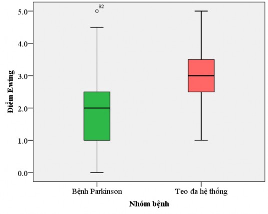
Figure 3.6 Comparison of Ewing scores between Parkinson's disease and multiple system atrophy
The Ewing score is calculated as the sum of the scores of 5 tests assessing cardiovascular autonomic function, including the deep breathing test, the orthostatic heart rate and blood pressure variability test, the Valsalva test, and the isometric contraction test.
The mean Ewing score in the Parkinson group was 2.02 ± 1.12. The mean Ewing score in the MSA group was 3.22 ± 0.96.
Comparison of 2 groups: There was a difference in the severity of autonomic dysfunction calculated by Ewing scale between Parkinson's disease and multiple system atrophy with p < 0.001 (t-test).