Pyramidon reagent method
In a test tube: 0.5 ml concentrated acetic acid, 2 ml urine, shake well; add 2 ml pyramidon 5% (in alcohol) and 0.5 ml H 2 O 2 3%. The mixture turns purple – positive reaction.
Phenolphthalein method ( Collo method )
In a test tube, add 3 ml of urine and 3 ml of Phenoltalein reagent; mix well and add 1 ml of 5% H 2 O 2 %. Red-purple contact ring – positive reaction
Reagents:
Phenolfthalein 2 g
KOH 20 g
Zinc powder 10 g
Distilled water 100 ml.
diagnostic meaning :
In urine there are red blood cells called hematuria, there is hemoglobin – hemoglobinuria and there is Mioglobin – Mioglobinuria. In diagnosis, attention is often paid to the first two forms.
Hematuria occurs when the kidneys, renal pelvis, renal tubules, bladder, or urethra are damaged or bleeding.
Renal hematuria: renal rupture, acute nephritis. Many infectious diseases cause bleeding: anthrax, bovine cholera, swine cholera, paratyphoid. Renal hematuria is dark urine, sediment with many blood clots, and renal epithelial cells.
Hematuria due to renal pelvis: renal pelvis stones, kidney worms, hemorrhagic pyelonephritis.
Bladder hematuria: cystitis, bladder stones, urethral ulcers, urethritis
bleeding… To differentiate, urine sediment test is needed.
To differentiate cases of urinary tract bleeding: collect 3 cups of urine at 3 times. Urine from the early, middle and late stages of urination, observe the color of the 3 cups.
If the color of the first cup is dark – due to bleeding in the urethra. The second cup is dark – bleeding in the bladder.
If all three cups are the same red color, then there is bleeding in the kidney or renal pelvis.
Hemoglobinuria is caused by excessive breakdown of red blood cells in the body and their release into the urine.
Differentiate between hematuria and hemoglobinuria
Hematuria Hematuria
Normal Eye Opaque Transparent
Packed red blood cells | Do not have | |
Check glasses | Intact red blood cells | Red blood cells rupture into pieces |
Filter multiple times | Discoloration | No color fading |
Maybe you are interested!
-

 Standard Teaching Hour Norms of Lecturers at Khxhnv University - Hanoi National University According to Each Position
 -
 Completing the training quality assurance system at the University of Economics, Vietnam National University, Hanoi - 16
 -
 SQL Database Administration - Hanoi University of Business and Technology - 14
 -
 Applying the process of caring for, preventing and treating diseases for breeding sows and piglets raised by their mothers at Thien Thuan Tuong Mineral Exploitation Joint Stock Company, Cam Pha, Quang Ninh province - 8
 -
 Marketing activities of James Hardiman Library, National University of Ireland, Galway and the possibility of application at the Information Center - Library of Vietnam National University, Hanoi - 1
Urine sugar test Heines method
Principle: if there is sugar, Cu ++ will be reduced to Cu + in Cu 2 O, brick or brown precipitate will form
dark yellow
This sensitive method is suitable for testing livestock urine, especially horse urine.
Heines reagent :
1. Dissolve 13.3 g of pure CuSO 4 in 400 ml of distilled water.
2. Dissolve 50 g KOH in 400 ml distilled water.
3. 15 ml pure glycerin in 200 ml distilled water.
Mix 1 and 2 together, stir well then pour in 3, shake well and store in a bottle with a cork for long-term use.
Testing
In a test tube: 3ml of Heines reagent, boil, then add 10 drops of urine test. If there is brick red precipitate: positive reaction.
Nylander method
Principle: in alkaline environment, bismuth nitrate is deoxidized by sugar to form bismuth oxygen or bismuth precipitates brown or black.
Reagents:
Sous nitrate bismuth 2.0 g.
Potassium – sodium tartrate 4.0g.
Potassium hydroxide (KOH) 100ml.
Dissolve and filter through filter paper, store in a colored glass bottle.
Consider :
In a test tube, add 3 - 5 ml of urine to be tested, 1 - 2 ml of Nylander urine,
Boil. The mixture turns dark brown: positive reaction.
Note: If the urine contains a lot of indican and other pigments, black precipitate will form when boiled. Therefore, before testing, add a little 25% HCl and bone powder, shake well, filter and discard. Urine containing a lot of mucus (muxin) can also give a false positive reaction.
Benedict Method
Similar to Heines method, but with the advantage that uric acid. Urate salts and organic residues are not capable of deoxidizing in Benedict's reagent. Benedict's method is widely used.
In Benedict 's test :
1) 173 g Sodium citrate (Na 2 C 6 H 5 O 7 .11H 2 O) and 90 g anhydrous Sodium carbonate (or 180 g crystalline Sodium carbonate) in 600 ml distilled water, heat gently, shake to dissolve, filter until dry and add distilled water to 850 ml.
2) 17.3 g CuSO 4 .5H 2 O in 100 ml distilled water, shake to dissolve then add distilled water to 150 ml.
Mix solution 1 with solution 2, shake well. Test
In a test tube: 1.5 ml Benedict's reagent and 3 drops of urine test. Shake well and heat for 2 minutes. Brick-colored precipitate: positive reaction.
The reaction results with Benedict's reagent can be used to calculate the amount of sugar in the urine.
Solution color
Result | Urine sugar concentration % | |
Constant | - | 0 |
Blue color without precipitation | | From 0.1 - 0.3 |
Blue precipitate | + | 0.5 |
Yellow | ++ | 1.0 |
Orange red | +++ | 1.5 |
Light red | ++++ | 2.0 and more |
diagnostic meaning :
The above testing methods detect glucose. In animal urine, in addition to glucose, there are also fructose, lactose, levulose, and pentose. Note that vitamin C, creatinine, and uric acid are also deoxidized like glucose, so they react positively with glucose tests.
Positive urinary tract tests are pathological and physiological.
Physiological urinary tract : Eating too much sugar, high blood sugar beyond the renal threshold. Cases: cattle are scared, excited, suddenly cold. Pregnant cattle urine contains lactose and this phenomenon disappears after the cattle gives birth after 2 - 3 weeks.
Pathological urinary tract : usually neurological diseases. Rabies, cerebral congestion, encephalomyelitis, poisoning cases (carbon monoxide poisoning , mercury poisoning, chloral hydrate poisoning). Some infectious diseases cause kidney damage and central nervous system stimulation. Chronic nephritis occurs in the urinary tract. Urinary tract in horses and dogs is a symptom of diabetes.
Urine bilirubin test
Urine with bilirubin is yellow, if there is a lot it is green, shaking has yellow-green foam. When the amount of bilirubin is small, it needs to be tested.
Method using nitric acid (HNO 3 ):
Filter the urine several times through a filter paper, then leave the filter paper on a box to evaporate until it is dry, the paper is white. Drop 1 drop of nitric acid in the middle of the paper, if concentric circles appear from the outside in: green, blue, purple, red, yellow. The green circle is due to bilirubin.
Method using 1% iodine alcohol :
In a test tube: add 1 ml of iodine alcohol, then slowly add 2 ml of urine tested on the iodine layer. If a blue contact ring is observed: positive reaction.
Method using BaCl2 :
Cut filter paper strips about 1.5 cm wide, dip them in saturated BaCl2 solution and dry them for long-term use.
Foucher's reagent: 25g acetic trichloride acid, 1g FeCl 3 in 100 ml distilled water.
Test: dip a piece of BaCl2 paper into the urine to be tested; take it out and dry it over an alcohol lamp flame or let it dry in the air. Drop 1 drop of Foucher's reagent onto the paper. Blue color appears: positive reaction.
Xinhop method :
Take 5 ml of urine, dilute with 5 ml of distilled water, then add 5 ml of 10% BaCl 2, shake well and filter through filter paper. Add 1 drop of fuming nitric acid to the residue on the filter paper.
If blue appears: positive reaction.
diagnostic meaning :
Bilirubin appears in urine when liver disease or bile duct obstruction occurs (see section “Liver examination”). In cattle, according to Roussow (1962), bilirubinuria occurs when serum bilirubin concentration exceeds 10 mg% and according to the author, cases are very rare. In buffalo, bilirubinuria occurs when serum cholebilirubin concentration exceeds 0.5 mg% (Ho Van Nam, Nguyen Kim Thanh, 1985).
Urine urobilinogen test
Urobilinogen in water includes four substances: urobilinogen, urobilin, stecobilinogen and stecobilin (Todorov, 1968).
Florens method :
Put into a test tube: 5 ml of urine to be tested, 4 drops of concentrated H 2 SO 4 to acidify and 3 ml of ethyl ether. Shake well, let stand for the mixture to stratify. Pipette the top ether into another test tube containing 3 ml of concentrated HCl. The next red ring appears: positive reaction. The more urobilinogen, the thicker the red ring.
Nhicop method :
The Neubauer method was improved by Nhicop based on the principle: urobilinogen combines with Ehrlich reagent to form a red compound and the amount of urobilinogen is determined by applying a certain amount of Ehrlich reagent to urine tubes of different dilutions.
Ehrlich reagent : Paradimethylaminobenzaldehyde: 2 g
HCl: 100 ml
Six equal test tubes, each tube 2 ml of distilled water and put into the first tube 2 ml of urine to test. Mix the first tube well, then draw 2 ml from the first tube to the second tube. Do the same with the second tube and draw 2 ml from the second tube to the third tube..., to the sixth tube, draw 2 ml and discard. Thus, the urine in the 6 tubes is diluted in the following ratios: 1: 2; 1: 4; 1: 8; 1: 16; 1:
32;...
Add 1ml of Ehrlich reagent to each tube, shake the tubes well and read the results after 5 minutes.
In cows, according to Nhicop: from the tube with urine dilution 1: 16 onwards, red color appears, positive reaction.
Urinary urobilinogen determination
Put into a test tube: 5 ml of urine, 5 drops of concentrated H 2 SO 4 to acidify and 2 ml of ethyl ether. Cover the test tube with a rubber tube and shake well. Leave for 5 minutes. Take 1 ml of the ether above into another test tube and add 1 ml of concentrated HCl. Shake well and leave for 10 minutes. Urobilinogen will turn into a red derivative and the red color will last for 12 hours or longer.
The Shali hemoglobin meter is redesigned by replacing the two side standard tubes with two
distilled water tube
How to measure: add the ether and acid solution above to the Shali tube up to the 5 mark (usually 5 - 6 drops), then dilute with distilled water until the red color disappears.
Calculation: Read the number corresponding to the concave surface of the solution in the Shali tube and divide by 5 to get the number of units. According to Mechiev, every 10 units (above) corresponds to 0.1 mg% urobilin.
For example, if the reading on the Shali tube is 40, the result is: 40: 5 = 8 units or 0.8 mg% The above simple quantitative method can be widely used in veterinary medicine.
Urine ketone test
Ketone bodies in urine usually have 3 substances:
In veterinary medicine, only qualitative tests are usually performed.
Lieben method
Lugol's reaction with acetone in an alkaline medium gives a yellow precipitate with an iodoform odor.
Test: put in a test tube: 10 ml of urine, a few drops of Lugol, a few drops of 10% KOH. Cloudy yellow precipitate, iodoform smell: positive reaction.
Lange method
In alkaline environment acetone combines with nitroferricyanic to form a purple-red mixture.
Test: in a test tube: 2 - 3 ml of urine, 5 drops of newly prepared saturated sodium nitroferricyanate and 0.5 ml of fuming acetone. Shake well, gently drop along the side of the tube and add 2 ml of ammonia solution. The contact ring appears purple-red: positive reaction.
Another way: put in a test tube 3 ml of urine, 1 ml of Sodium nitroferricyanate reagent (0.3 g sodium nitroferricyanate, 30 g ammonium nitrate and 80 ml distilled water).
Shake well, then slowly drop 2-3ml of concentrated ammonia water along the side of the test tube. The contact ring appears red: positive reaction.
meaning
The amount of ketones in healthy cattle is very small: one liter of horse urine has 0.38 - 3.56 mg%, cows: 0.2
– 2.4 mg ketones.
Increased ketones in the blood – ketonemia; and increased ketones in the urine – ketonuria.
Ketonuria is a symptom of lipid and carbohydrate metabolism disorders. In veterinary medicine, ketonuria is noted in dairy cows, as an important symptom of bovine ketonemia.
Ketonuria is also seen in postpartum paralysis, prolonged bed rest, and diabetes (Liabet).
Urine indican test Usually only qualitative test . Jaffe method
In a test tube: 5 ml of filtered, deproteinized urine, 5 ml of concentrated HCl, 1 drop of 2% potassium permanganate, 1 ml of chloroform. Close the test tube and shake well (20 times or more). Chloroform settles to the bottom of the test tube and turns blue. The reaction results are evaluated based on the color level. Purple reacts strongly (++++); dark blue is positive (+++); blue (-); pale blue (-).
Obermayer method
Obermayer reagent: FeCl 3 : 0.2g
Concentrated HCl: 100ml
Test: 3 ml of urine, 3 ml of Obermayer reagent in a test tube, shake well and heat slowly for 1 minute. Add 2 ml of chloroform, shake well and let stand for 3 minutes. The lower layer of the reaction is blue and depending on the light to dark blue color to estimate indican in urine.
The two methods for finding indican above are based on the principle that in an acidic environment, indican is oxidized by FeCl 3 or Potassium permanganate to form indigo red or indigo blue, which is soluble in chloroform.
diagnostic meaning :
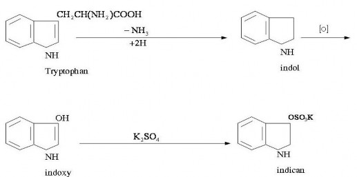
Indican is the final product of protein degradation in the intestine: for example, protein degradation produces tryptophan. Under the action of bacteria in the small intestine, tryptophan indole and skatol. In the liver, indole and skatol combine with H 2 SO 4 or glucuronic acid to form a non-toxic product - indican, which is excreted in the urine.
Indican is always present in urine, especially in horse urine.
Increased Indican is due to increased protein breakdown in the intestine: constipation, digestive disorders, intestinal obstruction, intestinal tuberculosis; abscesses, intestinal gangrene, severe uterine inflammation...
Indican reduced: diarrhea…
Urine chlorine test
Actually, NaCl should be tested because it is the main substance containing chlorine in urine.
Quantification of NaCl in urine
The simplest is to titrate with AgNO 3
AgNO 3 + 2NaCl (in urine) NaNO 3 + AgCl (white precipitate and the reaction is complete thanks to the color indicator K 2 CrO 4 ):
K 2 CrO 4 + 2AgNO 3 2KNO 3 + Ag 2 CrO 4 (black precipitate) Knowing the amount of AgNO 3 used, we can deduce the NaCl in the urine. Chemicals: 1. AgNO 3 solution : Pure AgNO 3 29.061g
Distilled water 1000 ml
Store in colored glass jar
Quantitative:
2. K2CrO4 10 %
3. NaHCO3 10 %
In a glass cup: 10 ml of urine, 40 ml of distilled water, 2 ml of 10% NaHCO 3 and 5 - 6 drops of K 2 CrO 4 color indicator. Shake well. Slowly add AgNO 3 (1), shake while adding, until black color appears.
Amount of NaCl in 1 day and night (g):
Number of ml of AgNO 3 used g 10
X Number of ml of urine of livestock
pee in a day and night
diagnostic meaning
The amount of NaCl in urine depends on many factors.
depends a lot on the nature of the food, the weather and the state of the body.
Decreased NaCl levels due to salt accumulation in the body: acute nephritis, acute infectious diseases, lobar pneumonia, osseous inflammatory processes.
Increased NaCl in urine: edema recedes, esophagitis recedes, and diseases with high fever are decreasing.
3. Urine sediment test
Making slides: centrifuge gently or let the sediment settle. Pipette a drop of urine sediment onto a glass slide and cover with a cover slip; add 1 drop of lugol to easily distinguish epithelial cells from white blood cells.
Can be smeared, fixed with methyl alcohol (methanol), stained with Giemxa dye or 1% methylene blue. Examine under microscope.
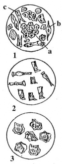
Organic matter :
1. Epidermal and dermal cells
a. The skin is dry, c. The skin is dry.
2. Epidermal cells
3. Bladder epidermal cells
Renal epithelial cells : round or square, with many small granules in the cytoplasm; round nucleus. Cells as large as leukocytes, exfoliated from renal glomeruli. There are many renal epithelial cells : acute nephritis .
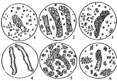
Renal pelvis and renal tubule epithelial cells : larger than renal epithelial cells , 3-4 times larger than leukocytes. Pear-shaped, oval-shaped cells. Due to pyelonephritis .
1. Traditional and wax patterns
2. Skin
3. Old customer service
4. The truth
5. Skin – Epidermis
6. Page
Bladder epithelial cells : diverse, fish scale-like , nucleus
round. The type of cells that come off from the deep layer of the bladder wall are smaller. There are many of these cells: cystitis.