2.3.2.3. Tests related to urine
- General urine analysis:
+ Indicated for patients who meet the criteria for choosing the disease after taking a medical history and having a clinical examination at the Urology Clinic, Emergency Department or other departments - Hue University of Medicine and Pharmacy Hospital upon admission.
+ Urine sample: collect about 5 ml of midstream urine into a plastic tube.
Maybe you are interested!
-
 Study on the treatment results of acute obstructive pyelonephritis due to ureteral stones - 23
 -
 Liver Function Tests in Child-Pugh Scale
 -
 Tests in Binary Logistic Regression Models
 -
 Research on Selecting Indicators and Characteristic Tests to Identify Models of High-Level Vietnamese Female Badminton Players
 -
 Techniques for Performing Autonomic Nerve Function Tests
Patients are instructed on how to collect urine to minimize contamination.
+ The test is performed at the Laboratory Department, Hue University of Medicine and Pharmacy Hospital. Using Roche dipsticks to read the results with the Japanese COBAS U411 automatic reader.
- Urine culture:
+ Indicated for patients who meet the criteria for choosing the disease after taking a medical history and having a clinical examination at the Urology Clinic, Emergency Department or other departments - Hue University of Medicine and Pharmacy Hospital upon admission.
+ Urine sample: collect about 8 - 10 ml of midstream urine into a sterile bottle, screw the cap tightly and send to the Microbiology Department as soon as possible, no later than 2 hours after collection. Patients are instructed on how to collect urine to limit contamination.
+ Urine culture is performed at the Department of Microbiology, Hue University of Medicine and Pharmacy Hospital. Urine culture results are usually available 48-72 hours after sample submission.
+ Procedure for urine culture
Shake and mix the urine well, collect urine with a quantitative inoculating stick with a volume of 0.001ml and streak it into CHROMagar Orientation medium: first, streak a straight line along the diameter of the agar plate, then use the inoculating stick to streak the entire inoculated urine to both sides in a zigzag pattern (the more even and thicker the better).
Incubate the streak culture medium in a 37ºC incubator for 28-24 hours, observe the colonies and count the number of colonies.
Identify bacteria based on physical, cultural, biological and chemical properties.
Bacterial quantification = number of colonies x 1,000 CFU/ml;
If >100,000 CFU/l ml then it is definitely pathogenic bacteria.
If <10,000 CFU/ml: no growth/no detection of bacteria causing urinary tract infection.
If 10,000 - 100,000 CFU/ml and accompanied by white blood cells or clear clinical symptoms, then think of pathogenic bacteria.
If ≥ 3 types of bacteria are present, it is considered contaminated.
2.3.2.4. Imaging related tests
- Unprepared urinary system X-ray
+ Indicated for patients who meet the criteria for disease selection and do not have an unprepared urinary system X-ray after taking a medical history and performing a clinical examination at the Urology Clinic, Emergency Department or other departments - Hue University of Medicine and Pharmacy Hospital upon admission.
+ The patient was filmed at the Department of Diagnostic Imaging - Hue University of Medicine and Pharmacy Hospital. The urinary system X-ray film was not prepared to meet the conditions for recording results with the limited standard of the film taken from the D11 vertebra to the lower edge of the pubic bone, balanced between both sides of the spine, the X-ray clearly shows the iliopsoas muscle, and the kidney shadow on both sides.
- Ultrasound of urinary system
+ Indicated for patients who meet the criteria for disease selection and have not had a urinary tract ultrasound after taking a medical history and performing a clinical examination at the clinic.
Urology, Emergency Department or other departments - Hue University of Medicine and Pharmacy Hospital upon admission
+ Research equipment: APLIO 500 ultrasound machine and Convex probe with frequency of 3.5-5 MHz at the Department of Diagnostic Imaging - Hue University of Medicine and Pharmacy Hospital.
+ Procedure: The patient lies on his back on the ultrasound table, with both arms raised above his head. Expose the entire abdomen, apply gel to the area to be examined. Use a 3.5MHz probe to examine the entire abdomen, then examine the kidneys, retroperitoneal space, bladder and entire ureter. The patient can be asked to inhale deeply, hold his breath to observe the kidneys more clearly. Use basic cross-sections such as coronal cross-section, longitudinal cross-section, and transverse cross-section to examine the entire kidney. Finally, examine the ureter, following the path of the ureter combined with rotating and scanning the probe for the best examination.
- CT scan of urinary system:
+ Indicated for patients who meet the criteria for disease selection and have images of ureteral stones on ultrasound and urinary system X-ray after taking medical history, clinical examination at the Urology Clinic, Emergency Department or other departments - Hue University of Medicine and Pharmacy Hospital upon admission.
+ Research means:
Siemens (Germany) 16-slice SOMATOM Scope CT scanner and efilm processing and storage software at the Department of Diagnostic Imaging - Hue University of Medicine and Pharmacy Hospital.
Intravenous contrast agent: low osmolarity, non-ionic contrast agent, osmolarity 600-900 mOsm/1kg H2O, Ultravist 300gI/ml. Contrast dose 1ml/kg, injected at 2ml/second with electric syringe.
+ Procedure:
The patient is explained in advance, the glomerular filtration rate is determined based on blood creatinine test before contrast injection.
Position: patient lying on back, hands above head.
Positioning image: Anteroposterior direction, length from diaphragm to pubic symphysis.
Examine the patient while holding their breath and inhaling.
Spiral cap 5/5mm.
Do not inject intravenous contrast
Intravenous contrast injection and imaging at different times:
Renal cortex phase (vascular phase): 30-40 seconds, examine the drug absorption in the renal cortex.
Renal medullary phase (renal tubular phase): 80-100 seconds, examines the absorption of contrast in the renal medulla, also considered the phase of contrast agent secretion into the renal tubules.
Excretory phase (late phase): 5-45 minutes depending on the level of obstruction, to see the urinary system's excretory pathway.
2.2.2.5. Initial empirical antibiotic therapy
Empiric antibiotic therapy was prescribed in all patients based on hospital-based bacterial sensitivity data after performing blood and urine culture tests [4].
2.2.2.6. Perform drainage of obstruction
Time of implementation: after patients are diagnosed with acute obstructive ureteral calculi (based on clinical symptoms and CLS index), the patient's condition can be treated with drainage of obstruction and has adequate equipment (operating room with enhanced brightness screen, ultrasound machine, etc.)
- Retrograde JJ ureteral catheter placement method via cystoscopy under fluoroscopy.
+ Indication: initial indication for patients in this study.
+ Equipment
Cystoscopy equipment from Karl – Storz, Germany (Hopkins 30-degree cystoscope, cysto 21F outer and inner sleeve, bridge for cystoscope).
Terumo Guide Wire Ureteral Catheter
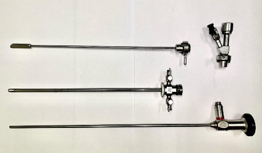
Figure 2.1. Cystoscopy instruments
(Source: taken at Hue University of Medicine and Pharmacy Hospital)
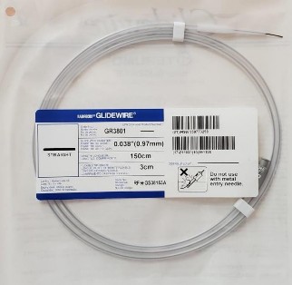
Figure 2.2. Terumo guide wire
(Source: taken at Hue University of Medicine and Pharmacy Hospital)
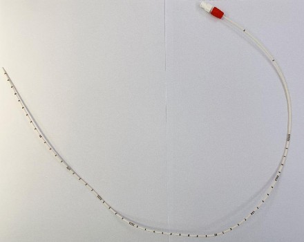
Figure 2.3. Ureteral catheter
(Source: taken at Hue University of Medicine and Pharmacy Hospital)
+ Procedure:
Patients and their families were clearly explained about the purpose, method, complications of retrograde JJ catheterization cystoscopy and unwanted symptoms when using JJ catheter.
Obstetric position as for conventional retrograde ureteroscopy.
Adjust the brightness of the monitor to the patient's position so that the bladder can be examined up to the renal pelvis.
Patients were analgesic with local anesthesia using 2% xylocaine gel injected directly into the urethra combined with intravenous analgesics (Fentanyl) or sedatives (Midazolam).
Place the 21F cystoscope into the bladder to find and identify the ureteral orifice on the side where the JJ catheter needs to be placed.
The guidewire is placed up to the renal pelvis past the stone obstruction under the control of the fluoroscopy monitor.
The ureteral catheter was then placed through the guidewire to the renal pelvis, the guidewire was withdrawn, and approximately 10 ml of urine above the obstructing stone was aspirated for urine culture.
Reposition the guidewire through the ureteral stone to the renal pelvis under the control of the fluoroscopy monitor and place the JJ ureteral catheter.
16F Foley catheter placement.
Figure 2.4. Obstetric position
(Source: taken at Hue University of Medicine and Pharmacy Hospital)
Figure 2.5. Ureteral stone (arrow) on X-ray film
(Source: taken at Hue University of Medicine and Pharmacy Hospital)
Figure 2.6. Ureteral stones (arrows) on CT scan
(Source: taken at Hue University of Medicine and Pharmacy Hospital)
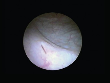
Figure 2.7. Right ureteral orifice